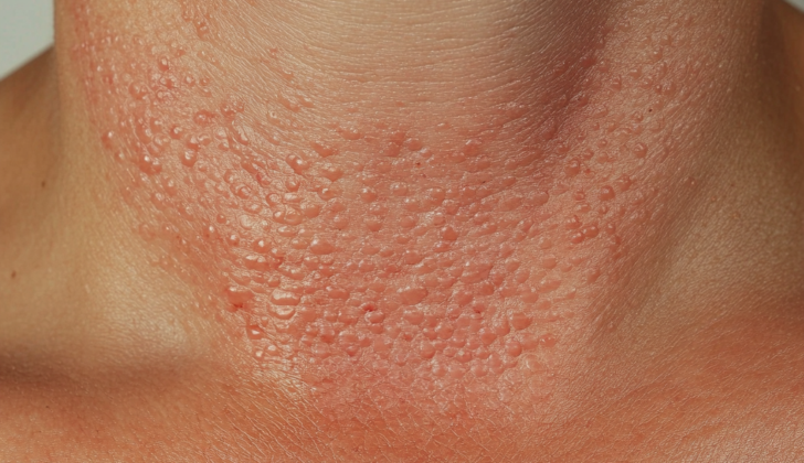
What is Tumid Lupus Erythematosus?
Cutaneous Lupus Erythematosus (CLE) is a skin condition related to lupus that shows up in three forms: acute (or sudden onset) CLE, subacute (or somewhat sudden) CLE, and chronic (or lasting) CLE. Chronic CLE also includes distinct types like tumid lupus erythematosus (TLE), discoid lupus erythematosus (DLE), chilblain lupus, and lupus panniculitis. These forms have unique appearances and microscopic characteristics. Moreover, it’s also possible for a patient to have more than one of these forms at the same time.
Even though TLE is currently labeled as a kind of CLE, it’s quite different from the other kinds. TLE rarely ties in with full-blown lupus, known as systemic lupus erythematosus (SLE). Due to this weak link with SLE and the fact that people with TLE often don’t show abnormal blood test results common in lupus, some believe that TLE might be a condition separate from lupus. Others speculate that TLE lies in the same category as lymphocytic infiltrate of Jessner and reticular erythematous mucinosis (REM) because they all share similar microscopic features.
TLE typically looks like red, swollen, hard, round patches or raised areas on the skin, without involvement of the topmost layer of the skin. If these characteristics are noticed and the top skin layer appears involved, it would suggest that the diagnosis might be DLE instead. TLE usually occurs on the face and trunk, and the skin changes respond well to sun protection, creams that lower skin inflammation, and malaria drugs.
What Causes Tumid Lupus Erythematosus?
So far, experts haven’t been able to pinpoint the exact cause of TLE, short for ‘Tumid Lupus Erythematosus’. However, they’ve noticed that certain things make it worse, such as exposure to ultraviolet (UV) rays from the sun. Some people think there might be a connection between TLE and autoimmune diseases, but the topic is still up for debate. When a doctor suspects an autoimmune disease, they might decide to perform some tests.
Some scientists believe that TLE could be due to the body’s immune system not working properly, specifically a part of the immune system called T cells. They think these cells might be suppressed, or not active enough.
Furthermore, TLE has been associated with unhealthy habits like smoking. Certain medications have also been pointed out as potential risk factors. These include drugs used to treat cancers or immunological disorders (tumor necrosis factor antagonists, monoclonal antibodies), high blood pressure meds (angiotensin-converting enzyme inhibitors), water pills (thiazide diuretics) and medication used to manage HIV (highly active antiretroviral therapy).
Risk Factors and Frequency for Tumid Lupus Erythematosus
CCLE, or Chronic Cutaneous Lupus Erythematosus, is generally seen more often in women. One type, TLE, or Tumid Lupus Erythematosus, is quite rare compared to another type known as DLE, or Discoid Lupus Erythematosus. We do not have data on how frequently TLE affects different races and ethnicities. The disease affects men and women almost equally. People usually start showing symptoms of TLE when they are around thirty to forty years old.

chest
Signs and Symptoms of Tumid Lupus Erythematosus
TLE, or Tumid Lupus Erythematosus, is a skin condition that doctors can diagnose by using a patient’s medical history as a crucial tool. If patients notice their skin getting worse with sun exposure, this might indicate TLE, though this symptom is not exclusive to this condition. For a thorough assessment, doctors will need to examine the patient’s entire body, paying special attention to the face, neck, chest, and back, as these are the most common areas for TLE.
The symptoms of TLE include swollen, usually circular skin patches that can range from reddish to purple. No changes to the uppermost layer of the skin usually occur with TLE — alterations like skin thinning, ulcers, plugging of hair follicles, scars, and changes in color suggest the presence of a different condition known as DLE (Discoid Lupus Erythematosus). However, it’s important to understand that TLE may sometimes show uncommon symptoms, which can include a pattern of skin changes that follow lines of cellular growth, swelling around the eyes, or hair loss that looks similar to a condition called alopecia areata.
It’s worth noting that TLE symptoms can last for days or weeks and have a tendency to occur repeatedly over time. While they can sometimes fade away on their own, some patients have reported that the symptoms return, especially during summer months.
Testing for Tumid Lupus Erythematosus
If a doctor suspects you might have a skin condition called cutaneous lupus, they might take a sample of your skin from an active, red area. This is done using a punch biopsy, a procedure that removes a small piece of skin. They typically remove a 4 mm piece if it’s from your body and 3 mm if it’s from more cosmetically important areas like your face. This biopsy should include the full thickness of the skin layer called the dermis.
The lab checks for certain patterns in the skin sample, like plentiful interstitial mucin and lymphocytes around blood vessels and hair follicles in the skin. If these specific signs are found, they would indicate a condition called tumid lupus.
If tumid lupus is diagnosed, it’s important to check if the lupus is affecting other parts of your body, despite it being less likely to do so. Your doctor will review your full medical history, check for swollen lymph nodes or arthritis, and order a number of laboratory tests. These include tests for antinuclear antibodies with specific subtypes, a urine test, a complete blood count, tests for certain chemical substances in your body, erythrocyte sedimentation rate (a type of blood test), C-reactive protein (which can indicate inflammation), levels of two proteins called C3 and C4, and antiphospholipid antibodies. They might also test for additional autoantibodies in order to further understand your condition.
If the biopsy shows signs consistent with lupus, but it’s not definitive, the skin sample can be sent for another test called direct immunofluorescence (DIF). The characteristic finding in cutaneous lupus is specific types of immune substances at the junction between the dermis and epidermis and around hair follicles. But in tumid lupus, DIF results often don’t show anything specific so it might not be as valuable.
An additional way of diagnosing this condition is by using phototesting, where your skin is exposed to UVA/UVB light. If the light exposure results in similar skin lesions, it would support a diagnosis of tumid lupus.
Treatment Options for Tumid Lupus Erythematosus
Firstly, for patients with localized TLE (Tumid Lupus Erythematosus), the best protection is avoiding direct exposure to sunlight and using topical or injected corticosteroids. It is recommended to regularly apply sunscreen with SPF 30 or higher, wear protective clothing, avoid peak sun exposure, and stop smoking.
The corticosteroids are directly applied to the skin or injected into the affected area. Usually, you would see an improvement in your skin’s condition after about two weeks of this treatment. Some side effects that can be seen from the use of these corticosteroids include thinning of the skin, stretch marks, lightening of the skin color, and small widened blood vessels.
If the condition doesn’t improve after a month, other treatments should be tried. Potent corticosteroids can be used for treating lesions on the trunk or extremities, while low potency corticosteroids can be used for facial treatment. Different types of corticosteroids are used according to the severity of the condition and where the lesions are located on the body.
It should be noted that topical calcineurin inhibitors such as tacrolimus and pimecrolimus could help improve skin conditions, while also reducing the need for topical corticosteroids. As these inhibitors don’t cause skin thinning, they are ideal for long-term use.
For patients with widespread TLE or those that don’t respond to topical treatments, the first-line treatment is usually antimalarial therapy. Hydroxychloroquine or chloroquine is initially used. These medications can cause skin depigmentation, resulting in a blue-gray discoloration on shins, palate, nails, or face which may be a permanent change. The most common side effects of these medications are stomach upset, leading many patients to stop the treatment. Other side effects can include issues with muscles and nerves, abnormal blood cell counts, and changes in vision. Therefore, regular eye exams are necessary while on this treatment.
TLE rarely doesn’t respond to antimalarial therapy, but if it happens, the next line of treatment involves drugs like methotrexate or mycophenolate mofetil. Side effects can include gastrointestinal upset, difficulty with breathing, suppressed bone marrow function, hair loss, and birth defects (in case of pregnancy). Healing with these drugs usually starts to be observed after two to three months of usage.
In rare cases, patients not responding to the above treatments might require third-line treatment options such as thalidomide and lenalidomide. Usage of these drugs might lead to adverse effects such as birth defects, nerve damage, blood clotting issues, and sleepiness. It’s necessary to taper down the doses of these drugs once the condition has improved to maintain remission.
What else can Tumid Lupus Erythematosus be?
When a doctor is diagnosing Tumid Lupus Erythematosus (TLE), it’s important to consider some other health conditions that may cause similar symptoms. These can include:
- Jessner’s lymphocytic infiltrate: Similar to TLE, it shows red bumps or lumps without scales on the upper back or face. Like in TLE, skin tissue tests can show a build-up of a type of protein. Some tests for immune system function may also come back positive.
- Polymorphic light eruption (PMLE): In PMLE, the skin becomes bumpy or changes color, and is itchy in sun-exposed areas. These symptoms often occur within a few hours after exposure to the sun and usually clear up quicker than TLE symptoms. Notably, PMLE doesn’t worsen with more sun exposure, unlike TLE.
- Reticular erythematous mucinosis: This condition causes red patches or plaques, usually on the upper back and chest. Like in TLE, skin tissue tests show protein build-up, but it’s more towards the skin’s surface, and immune cells are spread out more.
- Pseudolymphoma of the skin: This condition shows as red lumps in the skin layer, usually on the chest, arms, and face. They are not affected by light. Unlike TLE, skin tissue tests show presence of other types of immune cells.
- Granuloma faciale: This condition causes a purplish lump or plaque on the face, usually without any symptoms. What sets it apart from TLE is a specific finding on skin tissue test called the Grenz zone, which is an unaffected thin layer of skin tissue separating the inflamed lower skin layer from the upper skin layer.
For correct diagnosis, it’s important for the doctor to carefully take these possibilities into account and run the right tests.
What to expect with Tumid Lupus Erythematosus
Tumid Lupus Erythematosus is a condition that generally follows a harmless course. Most of the time, single lesions can heal by themselves, without needing any medication or treatments. However, it’s important to know that these lesions can return after a period without the disease.
Some patients have seen long-term remission, which means the condition disappears for an extended period. Interestingly, Tumid Lupus Erythematosus lesions tend to have a better outlook than lesions from discoid lupus erythematosus or subacute cutaneous lupus erythematosus, which are other types of Lupus.
Often, the lesions from Tumid Lupus Erythematosus can disappear by themselves without leaving any permanent marks or scars. This can happen within days or weeks, despite the potential for relapses. It’s also worth mentioning that over half of the patients were able to see their lesions disappear merely by protecting their skin from sun exposure, without experiencing any symptoms.
Possible Complications When Diagnosed with Tumid Lupus Erythematosus
It is uncommon for Systemic Lupus Erythematosus or discoid lupus erythematosus to be associated with each other. Some patients might experience vitamin D deficiency because they avoid exposure to the sun. Because of this, it’s recommended to check their vitamin D level with a 25-hydroxyvitamin D test. If a deficiency is found, a supplement of at least 400 IU of cholecalciferol (another form of vitamin D) is suggested.
Key Points:
- Association between Systemic Lupus Erythematosus or discoid lupus erythematosus is rare.
- Patients may develop Vitamin D deficiency due to lack of sun exposure.
- A 25-hydroxyvitamin D test can be used to verify Vitamin D levels.
- If Vitamin D is deficient, a supplement of 400 IU of cholecalciferol is suggested.
Preventing Tumid Lupus Erythematosus
Patients are strongly advised to protect themselves from sunlight by wearing protective clothing, reducing their overall sun exposure, and avoiding activities such as tanning, outdoor jobs, sunbathing, and traveling to places near the equator. It’s also recommended to apply sunscreen of at least SPF 50 twenty to thirty minutes before going outside. This kind of sunscreen protects against both UVA and UVB rays.
Patients are also encouraged to stop smoking because quitting has been shown to improve the condition of the affected area and the overall severity of TLE (a type of skin condition) when combined with antimalarial therapy.
For people whose condition does not respond to standard treatment, minimizing the use of medicines that increase sensitivity to sunlight is recommended. If a certain medication is causing TLE, stopping that medication can often resolve the issue. Patients should also be aware that new skin lesions can appear up to three weeks after being exposed to light.
Finally, patients should avoid touching or scratching TLE lesions, as this can lead to the Koebner phenomenon, a condition where skin lesions or rashes appear on areas of the skin that are injured or traumatized.


