What is Wallenberg Syndrome?
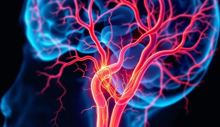
Wallenberg syndrome, also known as lateral medullary or posterior inferior cerebellar artery syndrome, was first detailed in 1895 by Adolf Wallenberg. This neurological disorder is caused by a blockage in either the vertebral artery or the posterior inferior cerebellar artery, which leads to an infarction (a blockage of blood supply) in a specific part of the brain called the lateral medulla oblongata. This damage to the brain results in various symptoms.
In terms of its anatomy, the posterior inferior cerebellar artery typically starts from the vertebral artery about 16 or 17mm below where the vertebral artery joins another artery at the base of the brain. The vertebral artery, originating from the subclavian artery (which comes directly off the aorta – the main artery of the body), is composed of four segments. The fourth segment gives off the posterior inferior cerebellar artery, which is the largest branch of the vertebral artery. This artery is divided into five distinct parts:
1. The anterior medullary segment starts at the beginning of the artery and ends near the inferior olive – a part of the brain located in the medulla oblongata.
From there it extends into the lateral medullary segment until it reaches the part of the brain where the nerves for swallowing and speakings (the glossopharyngeal, vagus, and accessory nerves) start. It then transforms into the tonsillomedullary segment until it starts to ascend (move upwards) towards the part of the brain that helps regulate the movement of cerebrospinal fluid. After that, it’s known as the telovelotonsillar segment, continuing until it leaves a particular groove in the brain. Finally, it enters its last segment, the cortical segment, which includes the final branches of the artery.
This artery gives rise to various other smaller arteries. It supplies blood to the medulla, the choroid plexus (a network of blood vessels), parts of the cerebellum, and other areas of the brain. Particularly, it provides the most blood supply to the choroid plexus on the roof and the median opening of the fourth ventricle – two crucial regions of the brain for the production and regulation of cerebrospinal fluid.
What Causes Wallenberg Syndrome?
Wallenberg syndrome typically results from a blockage of the main artery supplying blood to the brain, most commonly due to a buildup of fat and cholesterol, also called an atherothrombotic occlusion. After this, the blockage of the artery responsible for supplying blood to the lower part of the brain, known as the posterior inferior cerebellar artery (PICA), or rarely, the arteries to the medulla (part of the brainstem), is usually the next cause.
The importance of the PICA lies in its unique and complex anatomy and its involvement in a range of health issues, including stroke, bulging or bursting of the artery wall or aneurysm, pressure on the nerves or neurovascular compression syndrome (NVCS), abnormal connections between the arteries and veins in the brain or arteriovenous malformation (AVM), and brain tumors. Also, PICA is exposed to potential injury during several surgical procedures.
High blood pressure is often a major risk factor, along with smoking and diabetes. Another significant cause is when there is a tear inside the wall of the vertebral artery, a condition known as vertebral artery dissection. This condition can occur from neck manipulation or head injury, and from genetic disorders such as Marfan syndrome, Ehlers-Danlos syndrome, and fibromuscular dysplasia (an abnormal growth or narrowing of the medium-sized arteries in your body). Younger patients with Wallenberg syndrome commonly have a vertebral artery dissection.
Risk Factors and Frequency for Wallenberg Syndrome
Wallenberg syndrome is the most common type of stroke that affects the back part of the brain. In the United States, nearly 800,000 people suffer from a stroke each year. The vast majority, around 83%, are ischemic strokes, which are caused by blocked blood vessels. Roughly 20% of these ischemic strokes affect the back part of the brain. We can estimate that about half of these cases are Wallenberg syndrome, leading to over 60,000 new cases each year in the U.S.
This condition tends to be more common in men in their 60s. The main cause of Wallenberg syndrome is large artery blockages, which account for about 75% of the cases. Other causes include blockages originating from the heart, which cause about 17% of the cases, and vertebral dissection (a tear in the inner lining of the vertebral artery), which cause about 8% of the cases.
Signs and Symptoms of Wallenberg Syndrome
Wallenberg syndrome typically affects older patients with vascular risk factors and is a type of stroke syndrome. This condition often presents suddenly with symptoms like dizziness, vertigo, loss of balance, hoarse voice, and difficulty swallowing. These symptoms usually progress over a few hours or even days. It’s crucial to note that unlike most strokes, this syndrome usually doesn’t present with weakness, which can make it easy to miss or misdiagnose. For diagnosing Wallenberg syndrome, a detailed neurological exam is mostly relied upon. Although complete manifestation of the syndrome is rare, partial presentation is usually enough for diagnosis.
Generally, the diagnosis depends on detecting a combination of features typical of brainstem lesions and specific involvement of the posterolateral medulla region of the brainstem.
A patient with Wallenberg syndrome can present a variety of symptoms. These can affect either the side of the brain where the lesion is located or the opposite side.
On the side of the lesion, patients may experience:
- Vertigo with nystagmus (a condition that causes rapid, uncontrolled eye movement) and often accompanied by nausea, vomiting, and sometimes even persistent hiccups
- Voice hoarseness, slurred speech, and difficulty swallowing, usually accompanied by a reduced gag reflex
- Horner syndrome, which can cause drooping eyelids, constricted pupils, and lack of sweat on one side of the face
- Bias towards falling onto the side of the lesion, and one-sided loss of coordination and balance
- Pain, numbness, and impaired sensation on the face
- Impaired taste sensation
On the opposite side of the lesion, the syndrome may cause:
- Pain and impaired temperature perception in the arms and legs
- No or only slight weakness (this is because the fibers that carry motor signals are located towards the front)
Additionally, it might be useful to know that lesions located more towards the front of the brainstem often result in more severe difficulty swallowing and hoarseness. In contrast, lesions situated towards the back and side of the brainstem might present symptoms such as vertigo, loss of coordination and balance, nausea/vomiting, and Horner syndrome.
Testing for Wallenberg Syndrome
When it comes to diagnosing conditions that cause symptoms like dizziness, there are several things that doctors look at, as various conditions can present with similar symptoms. Some of these include:
1. Other causes of dizziness, such as acute labyrinthitis, which is an inflammation of the inner ear. Patients with this condition may be younger and not have any risk factors for stroke. Symptoms can include a certain type of unidirectional or rotating eye movement, called nystagmus, often accompanied by ringing in the ears, but no other brain symptoms. A particular eye movement test, called the head thrust test, can be used to differentiate a peripheral cause of vertigo, like acute labyrinthitis, from a central cause, like Wallenberg syndrome.
2. Hemorrhagic stroke: This type of stroke is less common, and usually involves a prominent headache.
3. Acute demyelination in multiple sclerosis (MS): Patients are generally much younger, more likely to be female, and have known history of the disease, which affects the protective covering of nerve fibers in the brain and spinal cord.
An acute relapse or attack of neuromyelitis optica, an autoimmune disorder that affects the spinal cord and optic nerves, can also cause similar symptoms. The patient is likely a young adult female, and the signs may suggest that more than one area of the brain or spinal cord is affected.
The diagnosis is usually suspected based on a clinical exam and a patient’s history of symptoms. The best diagnostic test to confirm if the symptoms were caused by a type of stroke that affects the lower part of the brain or side of the brainstem is an imaging test known as an MRI with diffusion-weighted imaging (DWI). However, it’s worth noting that up to 30% of patients who have had a stroke but show no disabling symptoms may not have observable lesions when this type of MRI scan is conducted. These patients are defined as DWI-negative stroke patients, who should start secondary prevention measures to prevent future strokes.
Additional diagnostic imaging, like a CT or MR angiogram, helps determine the place of the blockage in the blood vessels supplying the brain and can rule out more unusual causes like vertebral artery dissection.
An EKG can be useful to rule out any potential heart rhythm problems or acute heart conditions that could be related to the symptoms. Checking the blood for levels of key minerals (serum electrolytes) is also essential. If the patient is experiencing trouble swallowing (dysphagia) or speaking (dysarthria), it’s important to have them assessed by a speech-language pathologist before they eat or drink anything.
Treatment Options for Wallenberg Syndrome
The management of an acute ischemic stroke, where blood flow is blocked to the brain, relies on quick action. Rapid evaluation and starting treatment quickly can help reduce the size of the affected area (infarction) and prevent complications, improving the patient’s outcome and prognosis. That’s why it’s beneficial to manage these events in hospitals or stroke centers.
The management of a stroke usually involves certain steps. One approach is intravenous (IV) thrombolysis, a technique that uses a drug called tissue plasminogen activator (TPA) to dissolve the blood clot causing the stroke. This treatment is most effective if given within 3 to 4 1/2 hours of the onset of the stroke, and research shows it can improve the patient’s ability to function after the stroke by 30%. Some strokes, specifically those affecting the back of the brain (posterior circulation strokes), might have a longer treatment window.
Another approach is endovascular revascularization, a procedure using special devices to restore blood flow in the brain’s large vessels. This procedure has been shown to improve outcomes, especially in patients with large vessel intracranial occlusion, a severe type of stroke that can carry a poor prognosis without revascularization.
Patients are usually monitored in the intensive care unit (ICU) for 24 hours after IV thrombolysis. If ICU monitoring is not possible, the patient should be taken care of in a dedicated stroke unit. During the treatment, they will receive intravenous fluid to avoid dehydration but also to reduce the risk of swelling in the brain. Management of blood pressure is also important as the body’s natural regulation of blood pressure can be impaired in the affected areas of the brain. Speech therapy assessment might be necessary to test swallow function and prevent complications like aspiration pneumonia.
Other steps in management include deep vein thrombosis prophylaxis, which is a preventive measure against blood clots in the veins of the legs using pressure devices and blood-thinning medication. It’s also crucial to keep blood sugar within a healthy range, and antithrombotic therapy with aspirin may improve outcomes. Patients should also receive early physical and occupational therapy as part of a comprehensive rehabilitation plan.
Preventing a second stroke involves multiple steps tailored to each patient’s specific needs. These steps could involve surgery to remove a blockage in a carotid artery, oral anticoagulants for strokes caused by heart disease, blood-thinning medication such as aspirin, clopidogrel, or ASA/dipyridamole for other types of stroke, cholesterol-lowering medication, quitting smoking, controlling diabetes, maintaining a healthy blood pressure, and adopting a healthy diet and regular exercises. Adopting these multiple approaches can reduce the risk of subsequent stroke by up to 80%.
What else can Wallenberg Syndrome be?
The following medical conditions could be related to various symptoms:
- Chronic pain syndrome
- Lacunar stroke
- Middle cerebral artery stroke
- Migraine headache
- Multiple sclerosis
- Posterior reversible encephalopathy syndrome
- Subarachnoid hemorrhage
- Subdural hematoma
- Systemic lupus erythematosus
- Vertebrobasilar stroke
What to expect with Wallenberg Syndrome
In general, Wallenberg syndrome tends to have a better recovery outcome than most other types of stroke. Most patients are able to get back to their regular daily activities satisfactorily. The most common lingering issue is instability when walking.
Possible Complications When Diagnosed with Wallenberg Syndrome
Stroke syndromes can cause long-term disability and disrupt a patient’s daily life. Stroke syndromes, especially those related to the back of the brain, often lead to common complications like aspiration pneumonia (a lung infection caused by inhaled food, drink, or saliva), deep vein thrombosis (a blood clot in the deep veins), pulmonary embolism (a blockage in the lung’s main artery), and myocardial infarction (heart attack). Using breathing support like mechanical ventilation and dual-purpose feeding tubes can increase the risk of lung infections.
Here are the possible complications:
- Aspiration pneumonia
- Deep vein thrombosis
- Pulmonary embolism
- Myocardial infarction (heart attack)
- Possible lung infections due to the use of mechanical ventilation or dual-purpose feeding tubes
Recovery from Wallenberg Syndrome
Physiotherapy plays a crucial role in helping people with Wallenberg syndrome, a condition often caused by a stroke in an area of the brain called the lateral medulla, to regain their ability to function independently and return to their normal lives. Similar to other stroke treatments, the approach is individualized, tailored to suit each person’s specific needs and conditions.
The main objectives of any rehabilitation program following a stroke are to prevent any further complications, reduce symptoms, and increase the person’s independence and function. The goal is to make the training motivating, meaningful, engaging, and challenging to keep patients committed.
Rather than repeating the same set mechanical exercises, medical professionals often use a series of graded, real, and beneficial activities that are related to each patient’s lives. As a consequence, the treatment for Wallenberg syndrome varies based on what each patient struggles with. In many cases, patients may need therapies to improve their speech, language, and swallowing.
A certain kind of electrical muscle stimulation (NMES) has been approved by the US Food and Drug Administration. It helps specifically with a condition known as pharyngeal dysphagia – difficulty swallowing – and is typically administered by specially trained therapists such as speech and language pathologists and occupational therapists.
Physical therapy is also used to address challenges with balance, coordination, and movement, which are common symptoms of Wallenberg syndrome. The focus of these treatments is to train patients in specific day-to-day tasks, adapt to their environments, and retrain their motor skills to enhance their function and independence. Moreover, electrical stimulation has been shown to be effective in improving muscle strength and balance in people who have had a stroke. The current evidence supports the idea of using electrical stimulation to aid in motor recovery and is recommended to be incorporated at the beginning of the rehabilitation program.
Preventing Wallenberg Syndrome
It’s crucial that patients comprehend the long-term effects of their illness and consistently follow their prescribed medication and therapy routines. Doing so plays a significant role in enhancing their recovery process.


