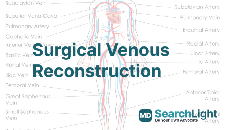
Overview of Surgical Venous Reconstruction
Chronic venous obstruction (CVO) is a medical condition where the veins get blocked. This can happen because of clotting following blood vessel injuries, trauma, medical procedures gone wrong, or removal of certain tumors affecting veins in areas known as the inferior vena cava and iliac veins.
Recently, there have been improvements in how we treat this type of blockage in the chronic state especially if it occurs in veins around the groin or abdomen, known as the iliofemoral or iliocaval veins. Now, a method called endovascular stenting is usually the go-to treatment, especially for non-dangerous or “benign” blockages. This method involves putting a small, flexible tube (stent) into the clogged vein with the aim to keep it open. This is typically used if initial, less invasive treatment methods haven’t worked.
However, if someone has severe symptoms from their CVO, and they can’t have or haven’t improved with this stenting procedure, they could be considered for surgical reconstruction of their affected veins. That involves an operation to repair or rebuild the blocked veins.
Anatomy and Physiology of Surgical Venous Reconstruction
Chronic venous insufficiency (CVI) is a condition that affects the drainage and flow of blood in the veins in the legs. Its symptoms can include swelling in the legs, skin changes, and the development of ulcers (open sores). These symptoms can occur due to conditions like high blood pressure in the veins, problems with the valves in the veins that stop blood from flowing backwards, or issues with the smaller ‘backup’ veins.
There are some risk factors for CVI that can’t be changed, such as being female, being younger than 50, and having a family history of venous disease. There are also risk factors that can potentially be changed, like being overweight, smoking, certain types of jobs, being pregnant, and using combined oral contraceptives (birth control pills).
In contrast to CVI, chronic venous obstruction (CVO) happens when there is a blockage in the veins. This blockage isn’t due to an issue with the walls of the vein or the valves within them, as is the case with CVI. CVO can be congenital (present at birth), primary (due to changes in the structure of the vein wall), or secondary (developing after a certain event). The causes of CVO can vary. They might include deep vein agenesis (veins that aren’t fully formed) or hypoplasia (veins that are smaller than normal), trauma, tumors, or certain medical procedures that involve the veins.
Why do People Need Surgical Venous Reconstruction
If you have Chronic Venous Outflow Obstruction (CVO), you may experience symptoms like leg swelling and pain, especially after exercise. These symptoms typically improve when you rest and elevate your leg. Other signs of CVO could include changes to the skin such as developing varicose veins, skin discoloration, eczema, tissue hardening (known as lipodermatosclerosis), and even open sores (ulcers). Your symptoms could be more severe if you have CVO compared to a similar condition called Chronic Venous Insufficiency (CVI). Depending on the condition of your blood flow, CVO could also potentially put your leg at risk.
Your doctor will want to understand your medical history to identify the potential causes of CVO. They’ll ask if anyone in your family has blood clotting disorders or venous issues, or if you’ve personally had a deep vein thrombosis (DVT) or inflammation of the veins (thrombophlebitis). They’ll be interested if you’ve had certain injuries or if you’ve been pregnant, as these are factors that can cause CVO. They’ll also check if you’re taking any medications, if you have history of abdominal surgeries or interventions, or even if certain aspects of your lifestyle or job could be contributing.
In examining you, the doctor will look out for signs like leg swelling, visible veins (even when lying down), skin discoloration, eczema, hardened tissue, and open sores on the legs. The side of the body where these symptoms appear will give clues as to the extent of the vein disorder. The doctor will also examine your abdomen and groin area to check for any obvious pressure on your veins, like a swollen abdomen or lymph nodes. They’ll look for varicose veins in the lower part of the body, like the groin and perineum, which could suggest vein issues elsewhere. The doctor may notice a network of veins, which could be a sign that blood flow is being redirected due to some kind of blockage. Lastly, varicose veins in the scrotum could indicate issues related to a vein entrapment condition (nutcracker syndrome), blockages in the main vein of the body (caval lesions), or even a type of kidney cancer (such as renal cell carcinoma).
When a Person Should Avoid Surgical Venous Reconstruction
Certain conditions make surgical repair of veins unsafe and these include:
If a person is extremely undernourished (severe malnutrition), their body may not have enough strength for the surgery and healing. Similarly, someone with a weakened immune system (immunocompromised state) might find it hard to fight off infections after surgery.
A person who is pregnant should avoid this surgery as it could affect the pregnancy. The surgery must also be avoided if there is an active infection in the area where the surgery would take place.
Another reason why surgical repair of the veins may not be performed is if a person has a blood condition (called uncorrected coagulopathy) that makes it difficult to stop bleeding.
Additionally, the surgery is not recommended for patients who are not medically stable, meaning their overall health is too poor to safely handle the procedure.
Equipment used for Surgical Venous Reconstruction
If you’re going to have a treatment for blocked or narrowed veins, the doctor will use a process called an endovascular procedure. They will use certain tools and machines to do this. Here’s a simplified list of what they use:
- An ultrasound – This is a painless test that uses sound waves to create images of your blood vessels.
- Access and working sheaths – These are tube-like structures that help guide the doctor’s instruments.
- Guide wires – These are thin, flexible wires that the doctor uses to navigate through your blood vessels.
- Diagnostic catheters – small, flexible tubes used to deliver therapy or diagnosis.
- Contrast material – a special dye that helps make your blood vessels more visible on an X-ray.
- Interventional devices – tools like balloons or stents to open up your blood vessel, or devices that remove blood clots.
- Intravascular ultrasound (IVUS) device – a special machine that uses sound waves to look at the inside of your blood vessels.
- Fluoroscopy unit – a machine that takes real-time, moving X-rays of your blood vessels.
- Personal protective equipment (PPE) – This includes things like lead aprons, vests, gloves, thyroid collars, and safety goggles to protect the doctor and healthcare team from radiation exposure.
If a surgical procedure to rebuild or reconstruct your veins is needed, your doctor will use standard surgical equipment. However, there are some tools specifically for this kind of surgery:
- Metzenbaum scissors – a type of surgical scissors used for cutting delicate tissues.
- Weitlaner retractor – a tool that keeps the wound or incision open during your operation so the surgeon can see and access the operative area.
- DeBakey and Gerald forceps – these are very slim pliers used to hold or manipulate tissues.
- Vessel loops – these are flexible loops used to temporarily control blood flow.
- DeBakey and Bulldog clamps – these are particular types of clamps used to temporarily close off blood vessels.
- Castro-Viejo needle driver – a tool for stitching or suturing the wound, specifically designed for delicate work.
- Nerve hook – tool used for dissecting nerves apart.
- Synthetic bypass graft – this is a man-made tube used to replace your vein in case your own veins are not suitable.
Who is needed to perform Surgical Venous Reconstruction?
To carry out a procedure on your veins, whether it’s minimally invasive or a traditional surgery, there’s a team of healthcare professionals that come together. This team usually includes:
1. An Interventionalist: This can be a vascular surgeon, interventional cardiologist, or interventional radiologist. These are specialist doctors who are trained to perform procedures on blood vessels.
2. A First Assistant: This person directly assists the main doctor during the procedure. They work hand-in-hand to ensure the procedure goes smoothly.
3. An Anesthesia Provider: This is the person responsible for making sure you don’t feel any pain during your procedure. They administer medications to help you sleep or numb the area where the surgery will take place.
4. A Circulating or Operating Room Nurse: This person doesn’t directly assist in the procedure. Instead, they make sure the operating room functions properly, by managing the surgical instruments and helping the rest of the team.
5. A Surgical Technician: Like an operating room nurse, they also provide important support. They help prepare the room and instruments before surgery, and assist during the procedure.
Preparing for Surgical Venous Reconstruction
For someone experiencing chronic venous insufficiency (CVI) or central venous obstruction (CVO), which means that blood is not flowing properly through the veins, doctors usually start with a Venous Duplex Ultrasound Scanning (VDUS). This test is safe, doesn’t hurt, and isn’t too costly. It can find out where the problem lies, the cause and seriousness of the issue by looking at venous visibility, compressibility, flow, and augmentation, which refers to whether the veins are visible, can be squeezed, have blood flowing within them, and if this flow can be boosted.
However, this method sometimes can’t thoroughly examine the upper iliac veins and the Inferior Vena Cave (IVC) – the two large veins that carry blood from the lower parts of your body towards the heart – especially in people with different body shapes and sizes, or too much gas in the intestines. If the results of the VDUS are normal, but doctors still suspect a patient to have CVI or CVO, another test called plethysmography has proven to be helpful. It assesses reflux (backward flow of blood), blockages and how well the calf muscle pumps – another important factor in maintaining good blood flow.
In cases where there might be a blockage in the IVC or upper iliac veins that is not clearly seen with VDUS, CT venography (CTV) and magnetic resonance venography (MRV) can be used. These tests use technology to take detailed pictures of the veins and help doctors see the venous anatomy, blockages, narrow points in veins and also identify whether there is something compressing the vein from the outside, like a tumor or a condition called May-Thurner syndrome.
Many consider intravascular ultrasonography (IVUS), despite being a bit more invasive and expensive, the best way to diagnose CVO. It uses an ultrasound device attached to a catheter that is inserted into the vein. It gives a clear, inside view of the vein and any area of interest. Some recent studies suggest that IVUS is actually better than a venography – another type of vein imaging test – in finding significant issues that other tests might miss.
Patients who are going for surgery might need an ascending and descending contrast venography. This can map both superficial (close to the surface of the body) and deep veins, evaluate valve function and identify the cause of faulty valves. It is carried out using a dye that enhances the visibility of veins. An ascending venography reveals obstructions and shows collateral venous circulation – the alternative paths that blood takes when the main veins are blocked. A descending venography, on the other hand, tests the valve function and identifies the causes of valve incompetence.
Lastly, doctors also use a technique known as ambulatory venous and arm-foot pressure measurements to determine the degree of venous hypertension – which is a condition where the pressure of blood in the veins is higher than normal. In some cases, doctors may recommend venous reconstruction if pressure differential – the difference in pressure between two points – is greater than 4mm Hg.
How is Surgical Venous Reconstruction performed
Percutaneous Venous Stenting
Percutaneous venous stenting is a widely used method for treating blockages in the veins of the lower body that aren’t responding to other treatments. It’s especially helpful for patients with CVI or CVO – conditions that cause issues with blood flow in the veins – who might not be good candidates for more traditional open surgeries. Open surgery, where an incision is made to access the veins, is still a choice in certain situations, such as when stenting isn’t suitable or when a cancerous tumor needs to be removed from the veins. Several factors can determine who is a good candidate for open surgery, including where and how long the blockage is, the cause of the blockage, how old the blockage is, and if the patient has an underlying cancer.
Sapheno-Popliteal Bypass, or the May-Husni Procedure
If there’s a blockage in the large veins of the thigh that doesn’t extend to the top of the leg (where the saphenofemoral junction (SFJ) is), a surgical procedure called a saphenopopliteal bypass, also known as the May-Husni procedure, can be performed. This involves creating a new connection between the large vein running down the back of the leg (the distal great saphenous vein or GSV) and another vein in the back of the knee (the distal popliteal vein) in order to reroute the blood flow around the blocked part. If the GSV on the same side as the blockage isn’t fit for the procedure, the one on the other side of the body or a vein from a donor can be used. Man-made replacements, such as those made of a material called polytetrafluoroethylene (PTFE), aren’t usually used in this area because they’re less effective.
Femoro-femoral Venous Bypass, or the Palma Procedure
For patients with a blockage in the veins of the thigh on one side, but normal flow on the other, a procedure called a femoro-femoral venous bypass (or the Palma procedure) can be performed. This involves using the large vein on the opposite side (the contralateral GSV) to connect the veins in both thighs, effectively rerouting the blood from the side with the blockage. Before the operation, an ultrasound scan is carried out to check if the GSV is fit to be used. A vein that is varicose (swollen) or less than 4mm in diameter might not be the best choice for this procedure.
Novel Valvular Repair Techniques
Treating CVI due to issues with the blood flow and the muscles around the veins in the lower legs is a bit more straightforward. But when CVI is caused by issues with the valves in the veins, it’s a little more challenging. New techniques are being developed that use devices made from pig aortic valve (part of a pig’s heart) attached to a metal frame. These can be surgically put into the veins and have shown promising results in treating CVI caused by faulty valves.
Possible Complications of Surgical Venous Reconstruction
There are often used treatment methods for chronic venous obstruction (CVO). Among these, doctors often prefer using endovascular procedures, which are treatments done inside your blood vessels. This method is generally considered safer and less intrusive than surgeries that require a larger cut.
Though endovascular procedures are helpful, they can also cause unique issues. Some of these issues can include:
* Bleeding or a build-up of blood (which is called a hematoma) at the spot where they entered your vein
* Rupture or tear of the vein due to the procedure used to widen the blood vessel (angioplasty)
* Puncture of the vein due to the tools used
* Blood clots in your veins
* A small metal tube called a stent moving to a different location or getting blocked
* An abnormal connection between an artery and a vein, which is known as an arteriovenous fistula
Surgeries that reconstruct your veins can also cause issues. Though these issues are similar to what could happen after most surgeries, they include bleeding, infections at the spot where the surgery was performed, and accidental injuries to nearby body parts.
What Else Should I Know About Surgical Venous Reconstruction?
In the past, issues with venous insufficiency (CVI) and venous occlusion (CVO) – conditions where the veins struggle to send blood from the legs back to the heart – were typically handled with non-invasive methods like compression treatments and exercises, or with more invasive surgical procedures. However, the use of endovascular techniques, which involve inserting a thin tube into the affected vein, has fast become the preferred choice for treating CVI and CVO.
Nowadays, surgical treatment is only suggested for those patients in whom the non-invasive or endovascular methods have not worked. Even though surgical procedures are being performed less often, doctors must still remain knowledgeable about these methods to ensure all treatment options are provided for the patients’ overall care.


