Overview of Catheter Management of Left Atrial Appendage Closure Devices
Atrial fibrillation, or AF, is a common irregular heartbeat condition that affects over 33.5 million people globally. The most serious problem that can occur because of this disease is a stroke, which is a key cause of disability in the U.S. If AF isn’t treated, it can increase stroke risk up to four or five times regardless of age. In addition, AF can also lead to the development of blood clots outside the brain, including in the aorta – the main blood vessel leading from the heart, as well as in the arteries of the digestive organs, kidneys, and limbs.
The rate of strokes due solely to AF increases with age, and can be as high as 23.5%. For those with AF, oral anticoagulants, or OACs – which are medicines that help prevent blood clots – are the best-known treatment. They have a clear role in preventing strokes. But they can’t be used in patients with a higher risk of bleeding. For these cases, an alternate treatment has been developed: left atrial appendage occlusion (LAAO), which involves closing off a small sac in the left upper chamber of the heart, where blood clots often form in people with AF.
Anatomy and Physiology of Catheter Management of Left Atrial Appendage Closure Devices
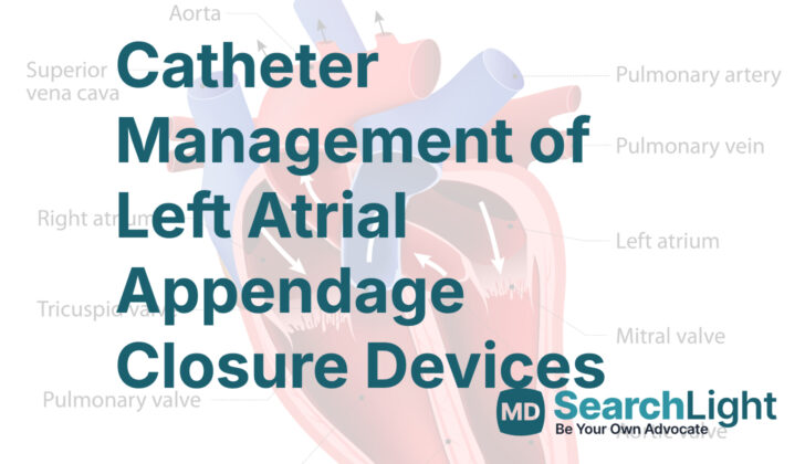
The left atrial appendage, or LAA, is a long, often bumpy tube connected to a specific part of the left side of your heart. It can look different in everyone, but for most people (about 70%), it bends or spirals. The LAA can be divided into three parts: the opening, the neck and the lobe area.
This area can reshape and enlarge over time, mainly in patients with a recurrent heart condition called atrial fibrillation or AF. In these cases, we can also see a decrease in pectinate muscle volume, these are the small muscles found inside the LAA. Using a special type of ultrasound of the heart, we see that between 5% to 15% of AF patients have blood clots, and an overwhelming majority of these clots are located in the LAA.
Under normal conditions, the LAA helps move blood in and out of its cavity when the heart is in a regular rhythm. But if there’s a problem with its contraction, like in the case of AF, blood may start to stagnate, leading to blood clots and the potential for these clots to travel to other parts of the body causing a blockage. The performance of the left side of the heart, which can be affected by AF, impacts how well the LAA can fill and empty of blood. In a healthy heart, when the heart chamber fills with blood it creates a suction effect which helps empty the LAA.
Why do People Need Catheter Management of Left Atrial Appendage Closure Devices
For patients afflicted with a condition known as atrial fibrillation (AF), which is an irregular or rapid heart rate, many health institutions consider a procedure called Left Atrial Appendage Closure (LAAC). This procedure is useful for AF patients who have a high risk of developing blood clots (known as a thromboembolic risk). This risk can sometimes be measured by a grading system called the CHADS2DS2-Vasc score—if this score is 3 or higher, or if the CHADS2 score is 2 or higher, LAAC is often considered.
LAAC may be recommended if these patients are unable to take long-term blood-thinning medications, known as oral anticoagulants (OACs), or if they have experienced blood clots even when using these medications properly. OACs might not be suitable if there’s a chance the patient may not take their medication as directed (termed therapeutic non-compliance) or if the patient can’t tolerate the medication due to side effects.
In 2019, the American College of Cardiology (ACC) and the American Heart Association (AHA) suggested that LAAC might be a suitable treatment for those with a high risk of stroke who can’t take blood-thinning medication long-term. The European Society of Cardiology (ESC) and the European Stroke Organization (ESO) gave similar advice in their 2016 guidelines.
The LAAC procedure works best when the height of the left atrial appendage (part of the heart) is larger than the width of its opening. Only one device, the Watchman (created by Boston Scientific), has been approved by the United States Food and Drug Administration (FDA) for reducing the chances of stroke in 2015. But this can only be used in patients with a high risk of stroke who can tolerate short-term blood thinners. Another device, the Lariat (from SentreHeart Inc), which is designed to help close off the left atrial appendage, is used off-label for LAAC in patients who can’t tolerate blood thinners and have a high risk of stroke. An ‘off-label’ use means it’s used in a manner not specified in the FDA’s approved labeling. However, there still isn’t much evidence to strongly support its use in these patients.
Lastly, if patients with AF are already undergoing heart surgery, there’s a consideration to also close off the left atrial appendage through surgery, although this suggestion isn’t as strongly supported as other treatments.
When a Person Should Avoid Catheter Management of Left Atrial Appendage Closure Devices
If a patient has a low risk for a stroke – meaning they have a CHADS2DS2-Vasc score of less than 3 or a CHADS2 score of less than 2 – doctors generally advise against a procedure called Left Atrial Appendage Closure (LAAC). Another reason a person might not be a good candidate for this procedure is if they have certain types of heart disease, like mitral stenosis.
This procedure is also not recommended for people with a clot or a tumor in the left part of the heart, an active infection, a hole in the wall between the two upper chambers of the heart (atrial septal defect), or if they have a device due to a condition called patent foramen ovale (a hole that didn’t close as it should after birth).
People who need to take blood thinners for a long time – maybe because they have a prosthetic valve, blood clots in the lungs (pulmonary embolism), blood clots in the deep veins of the body (deep vein thrombosis), or blood clots in the left side of the heart – should not have this procedure either.
Another reason a person might not be able to have Left Atrial Appendage Closure is if the depth of the Left Atrial Appendage (LAA – a small pouch in the upper part of the heart) is considerably shorter than the width of the opening to the LAA. As a rule, this procedure is also not advisable for patients who can’t tolerate a short course of oral blood thinners.
Equipment used for Catheter Management of Left Atrial Appendage Closure Devices
In 2001, a device known as PLAATO was used for the first time in human medical procedures by Dr. Sievert. PLAATO was made by Appriva Medical and was crafted from a self-expanding metal called nitinol. It also had a non-thrombogenic polytetrafluoroethylene coating (simply, a material that prevents blood clots) and just like a tree has roots, it had anchors along three struts to help the device secure itself in the left atrial appendage (LAA, a small pouch in the heart). It came in various sizes. Unfortunately, despite good results from clinical trials, the device was withdrawn from the market in 2006.
The Watchman device by Boston Scientific is currently the only one of its kind that’s been tested through randomized clinical trials. It’s made of a semi-spherical frame with a fabric cap on one end. The size of this device is determined by the diameter of its widest portion. It’s implanted into the left atrial appendage (LAA) of the heart, and its size must be slightly larger than the LAA itself. If the implant is too deep or the size isn’t right, the device can be partially or fully recaptured and redeployed. It’s currently approved for use in the U.S. and for sale in the European Economic Area.
The Watchman FLX, another device by Boston Scientific, had total coverage by a polyethylene membrane to reduce leaks. Unfortunately, it was removed from the European market due to increased implant issues.
Next we come to AMPLATZER Cardiac Plug (ACP) and the Amulet device, both by Abbott Vascular. The ACP is a self-expanding mesh device with a fabric disk and a connected lobe. In simple terms, it’s a plug that can adapt well to shorter LAAs. If needed, this device can be recaptured and re-positioned. The Amulet, on the other hand, is an upgraded model of ACP with added enhancements that facilitate implantation and reduce complications during the procedure. It’s also available in a more extensive size range and provides better stability in the heart.
The WaveCrest device by Biosense Webster, is a single-lobe nitinol device with anchor points to secure it, and it can be manipulated independently during the procedure for an adequate seal. It was given approval in Europe in 2013.
The Occlutech LAA Occluder device, developed in Sweden, has a conical shape and captures the distal margin through closed loops. It also uses a polyurethane cover to facilitate sealing and endothelization, which is the process of forming a new layer of cells in the heart. The device got approval in Europe in 2016.
The LAmbre LAA Closure System is made in China and it comes in two models: a standard one and a special one for individual heart anatomy needs. It has a unique double stabilization mechanism to secure the device and can adapt to a wide range of LAAs, and it’s possible to reposition or recapture the device during the procedure. This device got European approval in 2016.
Lastly, the Ultraseal LAA closure device by Cardia Inc, is made of two sections: a distal bulb for anchoring and a proximal sail for occlusion of the LAA. It is designed to adjust to varying shapes and angles of the ostium (opening) of the LAA. It also has multiple stabilizing hooks to prevent being dislodged during the procedure.
Who is needed to perform Catheter Management of Left Atrial Appendage Closure Devices?
The procedure is done in a specific type of medical lab meant for heart-related procedures. The main members of the team involved in your procedure will be an anesthesiologist (a doctor who administrates drugs for sleep or pain), an imager (a healthcare professional who takes and processes the images of your heart), and the implanter (the doctor who carries out the actual procedure).
You will be given a powerful sedative to make sure you are asleep and can’t move during the procedure. This is to keep your airway open, as well as to prevent unexpected movements that could potentially risk puncturing your heart when the doctors are working on the heart’s left atrial appendage (LAA) – a small pouch in one of the heart’s four chambers.
The X-ray machine (called fluoroscopy) is placed on the operator’s left-side so they have more space to work. Your echocardiographer (a specialist who uses sound waves to develop pictures of the heart) is seated at the head of the table. The echocardiographer and anesthesiologist are given special protective gear to shield them from radiation.
The operator (the main doctor conducting your procedure) can see images of your heart in real-time on an ultrasound machine that is connected to a mechanical arm.
Preparing for Catheter Management of Left Atrial Appendage Closure Devices
Doctors use a medical imaging method known as a transesophageal echocardiogram (TEE) before a procedure. This helps them understand if the patient is a good candidate for the procedure by checking the size and shape of the left atrial appendage (LAA), which is a small, ear-shaped sack in the muscle wall of the top left chamber of your heart. Important things they are looking for include the shape and size of the opening, the width of the area where they will place the device, the length of the LAA, and the number, shape, and position of any lobes (sections of an organ).
The doctors also check where the device will be positioned within the left atrial appendage, this is what they call the “landing zone”. If the neck of the LAA is bigger than usual (more than 26 mm), or the shape of the LAA is complicated, the doctors might consider using other imaging methods like a CT scan or MRI. Since the devices have specific size limits, good planning can avoid having to stop the procedure after the patient has already been given general anesthesia.
Factors like the amount of pressure in the left atrium and whether the heart rhythm is regular or irregular can affect the size of the LAA. To get the most accurate measurement, some doctors prefer to take these measurements during the procedure. They might infuse saline to create a pressure of more than 10 mm Hg in the left atrium. After the procedure, to make sure there’s no fluid around the heart (pericardial effusion) or if the device has moved to an unwanted location (embolization), many centers also perform a chest x-ray and a special test called a TTE.
To make sure the LAA is completely sealed and there’s no blood clot, doctors recommend doing a TEE after 45 days. After the procedure, patients might take different medications to prevent blood clots. These might include warfarin, combined therapy with two antiplatelet drugs (DAPT), direct oral anticoagulants (DOAC), aspirin alone, or even no therapy. The studies show that using DAPT or DOAC doesn’t significantly increase the chances of device thrombus (clot), stroke, or bleeding compared with standard warfarin therapy.
How is Catheter Management of Left Atrial Appendage Closure Devices performed
The following procedure describes how a particular device is implanted to close off the left atrial appendage (LAA), a small pouch in the heart, from the rest of the heart. This is done to prevent blood clots from escaping the LAA and potentially causing a stroke. This surgery is usually done under general anesthesia, meaning you’ll be asleep and won’t feel any pain.
Right before the procedure, you will likely be given antibiotics to prevent infection. The surgeon will gain access to your heart by making a small incision in the femoral vein, located in your groin. A narrow tube will then be threaded through this incision to your heart. The exact spot for this tube to pass through depends on the orientation and size of the LAA which varies patient to patient.
The type of device and its size depends on the size and form of your LAA. They come in different shapes and sizes to fit different patients. The surgeon will decide which device is best for you. Ultrasound and x-ray (fluoroscopy) will be used to monitor the device as it’s being placed in the LAA. Once the device is in place, the surgeon will do a ‘tug test’ to make sure it’s secure. Color Doppler imaging, a special type of ultrasound that can see blood flow, will be used to make sure the device is completely sealing off the LAA.
Depending on the specifics of your LAA, other devices might be better options. The “Watchman” device is chosen based on the size of LAA using a combination of ultrasound and x-ray. The device is preloaded onto a delivery system and guided to the LAA using a delivery cable. The device is then released once the surgeon is happy with the positioning.
Amplatzer Cardiac Plug, and Amulet devices are delivered to the LAA from the right femoral vein, just as one might deliver the Watchman device.
The Lariat device is different from the others because it requires an intact pericardium, which is the sac that the heart sits in. This means that patients with previous cardiac surgery or pericarditis may not be candidates for this device. The device is delivered with the help of a magnet-tipped guidewire which is more technical than the other devices but has its advantages.
The WaveCrest device is positioned to block the LAA and is tested to make sure it’s doing its job. The anchors are then placed at the edge of the device. Similar to the other devices, a ‘tug test’ is done before release.
The specific device and procedure will vary based on your individual anatomy and condition. Your doctor will discuss which options are right for you.
Possible Complications of Catheter Management of Left Atrial Appendage Closure Devices
The most frequent issues that might emerge after a specific heart procedure are: a build-up of fluid around the heart (also known as pericardial effusion), or sudden excessive pressure on the heart (cardiac tamponade) demanding treatment; formation of clots on the device used; the device moving from its original placement necessitating removal; a lasting hole in the wall separating the heart chambers (persistent atrial septal defect, or ASD); a break or tear in the heart; and stroke related to the procedure.
The possibility of a pericardial effusion or cardiac tamponade ranges from around 1.2% to 5%, and in most patients, a treatment known as pericardiocentesis, which uses a needle to remove the extra fluid, can be executed. Also, there can be a chance, although high at around 3.5%, the device could relocate to another part of the body, which might require surgical removal or readjustment. Nevertheless, the likelihood noticeably decreases when experienced professionals are handling the procedure (to less than 1%).
The device may sometimes contribute to clot formation, which can occur in up to 14% of patients. Usually, this can be managed by lengthening the course of anticoagulant medication, a type of drug, which helps prevent blood clots, with an effectiveness rate close to 95% (median treatment duration of nearly 45 days). The good news is, most patients experiencing this do not have any symptoms, and the overall risk of neurological events or problems related to clotting from the device is low (0.28%).
Persistent ASD may occur in around 11% of people after six months and in 7% of people a year following the procedure. Most often, the ASD is small and doesn’t need any extra treatment. Heart perforations, which are like tears, are rare and happen in up to 0.4% of patients. These usually require a surgical intervention. As well, up to 1.1% of patients might experience a stroke related to the procedure needing management.
Less frequent complications include fatalities related to procedure and vascular issues such as a hematoma (an abnormal collection of blood outside blood vessels), bleeding, and AV fistula formation (an abnormal connection between an artery and vein).
What Else Should I Know About Catheter Management of Left Atrial Appendage Closure Devices?
The Watchman device safety and effectiveness in patients with atrial fibrillation (an irregular heart rhythm) has been studied extensively. Two large clinical trials and several registries have shown consistent and positive results in terms of successful procedures and reduced complication rates. One significant trial, called PROTECT-AF, showed a failure rate of about 9% and a complication rate of about 8.7%. Most complications were due to an excess of fluid in the space around the heart (pericardial effusion). However, less harmful strokes occurred in the group who received the WATCHMAN device compared to the medication group.
The subsequent PREVAIL trial involved more patients, including those at high risk and doctors new to the procedure. To put it simply, it compared patients eligible for the WATCHMAN device with patients who could receive a blood-thinning warfarin medication. The results showed that major complications happened less often in the Watchman group, and the device proved to be just as effective at preventing stroke or a blood clot in the body.
Other studies have shown similar results, demonstrating success rates of over 95%, and very low rates of serious side effects within a week of the procedure.
Another device called the ACP was also studied. It showed a high success rate and a few serious side effects.
All these devices were designed to treat atrial fibrillation, a common heart rhythm disorder. But some patients with certain anatomical features around their heart just couldn’t use them. For these patients, the Lariat device is a safe alternative, even though there are some safety concerns. Studies of the Lariat device have shown success rates of about 94%, with a few major complications.
In conclusion, all the devices mentioned above have demonstrated high effectiveness and safety when used to treat atrial fibrillation. However, as with all medical procedures, there may be some risks, so it’s essential to discuss these with your doctor.


