What is Diving Gas Embolism?
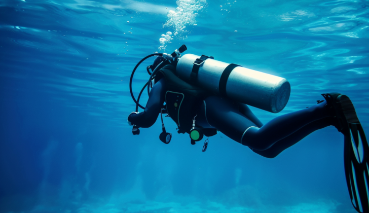
Diving with any kind of underwater breathing device means the diver breathes in compressed gas at pressures above what’s normal at the surface. Since seawater is denser than air, the pressure it exerts is equal to the pressure from one “atmosphere” (a measure of air pressure) at about 33 feet under the water. This means the pressure on a diver doubles in the first 33 feet under water.
When diving, a rule called Boyle’s Law comes into play. It says that as pressure decreases, volume increases in the same proportions. This means if a diver holds his breath while going up from 33 feet underwater, his lung volume would double once they’re back at the surface – if that was physically possible without harming the lungs. However, conditions like having overly reactive airways or certain anatomical quirks, like bulges in the lungs, can trap air inside the lungs if a diver holds their breath.
If a diver ascends even just 3 feet, it can lead to an overpressurization that’s strong enough to burst small air sacs in the lungs and release gas into nearby tissues and blood vessels. This condition is known as “pulmonary overinflation syndrome” and can result in injuries from overexpansion, such as having air trapped in the chest cavity, collapsed lungs, air pockets under the skin, or gas bubbles in arteries.
What Causes Diving Gas Embolism?
An arterial gas embolism is a rare condition that occurs in diving and in cases of sudden pressure changes such as in commercial planes, military aircraft, or spaceships. However, it’s more common that gas can enter the arteries during medical procedures like CPR, venous access, surgical procedures or needle biopsies. Basically, any time a medical operation involves the blood vessels, there’s a possibility that gas could be introduced.
Small amounts of gas, typically less than 30 cc, that enters the veins are usually filtered by the lungs and won’t show symptoms most of the time. However, if gas enters the arteries, it can become trapped in smaller arteries, resulting in symptoms similar to damage to the end organ. When gas enters the brain arteries, it can cause symptoms similar to a stroke and can make a person unconscious.
Risk Factors and Frequency for Diving Gas Embolism
Decompression illness, also known as DCI, is a term used to describe a variety of health problems connected with changes in pressure. It has two main types:
- Decompression sickness, or DCS, is a group of health issues caused by the formation of bubbles due to gas being dissolved in the body.
- Overexpansion injuries, included in these are arterial gas embolisms, or AGE, which occur when any type of gas in the body expands directly due to the effects of pressure change (known as Boyle’s Law).
These types of injuries are really rare and mostly related to diving accidents. Overall, the rate of diving-related health problems is between 1 to 3 for every 10,000 dives. Arterial gas embolism is even less common, happening in fewer than 1 out of every 100,000 dives.
There’s also a case of gas embolism that can occur due to medical procedures, which could introduce gas into the body’s arteries. This type is treated similarly to diving embolisms, but is not generally associated with lung damage as it typically doesn’t involve pressure changes similar to diving.
Very rarely, overexpansion injuries can happen from explosions or sudden decompression in planes or spacecrafts.
Signs and Symptoms of Diving Gas Embolism
Gas embolism, a potentially deadly condition, often has links to sudden decreases in environmental pressure or recent invasive medical procedures. Patients at high risk include divers, particularly those who ascend too quickly or lose control of buoyancy and hold their breath. Certain medical procedures, such as the insertion or removal of a central line, cardiac catheterization, vascular operations, and different surgical interventions, can also cause gas embolism. In some rare instances, gas embolism has occurred from ingesting hydrogen peroxide, inhaling directly from a helium tank, or during specific intimate activities during pregnancy.
Those suffering from a gas embolism typically lose consciousness within 10 minutes from the onset of the condition or from surfacing after a dive. The onset can be characterized by mixed neurological symptoms. It’s important to note that these symptoms may not connect to a singular cause which makes diagnosing the gas embolism a pretty complex process. Usual signs include physical weakness or even paralysis. However, it’s critical for healthcare providers to conduct comprehensive neurological examinations, including tests for cerebellar function, as the symptoms could be subtle and easily overlooked.
Testing for Diving Gas Embolism
Detecting a cerebral arterial gas embolism, which is a gas bubble in the brain’s blood vessels, relies on a patient’s medical history and symptoms. For example, situations such as taking out a central venous line (a tube inserted into a large vein) or coming up from a deep dive could introduce gas into the bloodstream. If a person faints or shows neurological symptoms within 10 minutes of such an event, this gives strong hints of an embolism.
While a chest x-ray or a head CT scan (brain imaging) can provide helpful information, these tests aren’t always necessary if the history and symptoms clearly indicate a gas embolism. Such imaging can help rule out other conditions like a collapsed lung, or if the cause of the patient’s symptoms isn’t initially clear. A simple finger-prick blood sugar test is also recommended, as similar symptoms can occur with both low blood sugar and gas embolisms. This test won’t delay treatment.
One important thing to remember is that divers, who are at significant risk of gas embolisms, can also develop other medical conditions. So, when examining a patient, it’s crucial to consider other possible diagnoses. However, if a gas embolism is suspected, treatment shouldn’t be delayed. Even if a head CT scan looks normal, it doesn’t rule out a gas embolism since the gas bubbles usually disappear quickly.
If gas is seen on a head CT, this is definitive proof of a cerebral arterial gas embolism, and the patient needs immediate treatment. Nowadays, it’s known that seeing gas on a scan indicates a large amount of gas in the body, which is often linked to severe disease and higher risk of death.
Large amounts of gas frequently cause multiple emboli, visible on CT or MRI as irregular patterns. Even in the absence of visible gas on a CT scan, it doesn’t mean a gas embolism hasn’t occurred. MRI may show no changes at all, or widespread areas of tissue damage with swelling. The inconsistency between imaging results and confirmed gas emboli remains an unresolved mystery in medical research.
Treatment Options for Diving Gas Embolism
The cerebral gas embolism treatment involves rapidly increasing pressure with pure oxygen. As a first-aid measure, patients are given 100% oxygen with a mask until recompression. Patients who are unstable or have a serious decline in consciousness might need a tube put into their windpipe to help with breathing, especially if the pressure chamber can support a ventilator. It’s crucial to consult with a hyperbaric physician as soon as possible, especially for critical cases.
For fluid replacement, non-dextrose-containing solutions are used. It’s also important to note that while early animal studies recommended the use of Lidocaine via injection, recent studies did not find it beneficial. Also, aspirin is not proven to be beneficial for this condition. The treatment often requires multiple hyperbaric sessions (3 to 5 or more) until the patient’s condition changes significantly. But usually, there is an immediate improvement with recompression. However, the longer the wait for treatment, the less likely there will be an immediate improvement.
In rare cases, a gas embolism could occur together with a blood-clot, which leads to mixed symptoms. There’s insufficient literature to guide the treatment of this situation. In some instances, outcomes have worsened when acute stroke was treated with hyperbaric therapy. These cases should be treated with utmost care by the hyperbaric physician, even involving a neurologist if necessary.
Though a mask providing high-concentration oxygen could initially improve or even resolve symptoms, it’s not sufficient for treating gas embolisms because symptoms often return.
For divers suspected of having a gas embolism, they should be given high-concentration oxygen and quickly taken to an emergency department equipped to diagnose and treat serious neurological injuries. Previously recommended positions for transportation, such as laying the patient downwards or on their side, are no longer advisable.
The widely accepted hyperbaric oxygen treatment is the US Navy Treatment Table 6, which may be switched to Table 6A if no improvement is seen in the first 10 minutes. This is most effective within two hours from the start of symptoms. Outside of this two-hour timeframe, running an extended US Navy Table 6 back-to-back is considered adequate. The reason behind this protocol is that any gas has likely moved through the bloodstream, so you don’t need to try to “crush” the bubbles with a rapid increase in pressure up to 165 feet below sea level. This also reduces risks for the caregiver and the patient. Confusion regarding diagnosis or achieving a satisfactory result with initial measures might encourage longer Table 6 treatments. For medically caused cerebral gas embolism, without inhaling compressed gas, compression to 165 feet below sea level might be unnecessary as these patients usually don’t have excess gas saturation in their bodies.
What else can Diving Gas Embolism be?
Cerebral arterial gas embolism, a condition harmful to divers, can sometimes be confused with a stroke; this is because both conditions show similar symptoms such as loss of consciousness, altered mental status, and crossed neurological deficits. In cases where a stroke is thought to have occurred after diving, physicians may consider a quick, non-contrast CT scan of the brain to diagnose it, but it’s crucial not to delay the diver’s recompression – a treatment to reverse decompression sickness – for more than a few minutes.
Low blood sugar, or hypoglycemia, can also cause the same symptoms, and is relatively common, so it’s a good idea for the doctor to check the blood glucose level as it won’t delay recompression. Sometimes, rare neurological disorders may also mimic the symptoms of a gas embolism, but these aren’t likely to appear instantly after a dive, so they can typically be ruled out until recompression has happened.
Venous gas emboli (VGE), or gas bubbles in the veins, are often found in divers after a dive. If these bubbles get into the arterial circulation through a shunt (a passage or a hole), they can block the blood flow to tissues and cause symptoms similar to those of a stroke, including severe neurological symptoms soon after surfacing. There are cases when the symptoms caused by these bubbles might not be the same as those of an arterial gas embolism due to lung inflation. However, it can be hard to tell these conditions apart at first. The treatment approach is similar for both, so early recompression is considered a priority.
Nonetheless, correct diagnosis becomes crucial when considering further tests and assessing the diver’s fitness to continue diving. For instance, a diver who experiences severe neurological decompression sickness after a normal dive might need to be checked for a patent foramen ovale (a hole in the heart) while someone who suffers an inexplicable arterial gas embolism might need a radiographic examination of the lungs.
What to expect with Diving Gas Embolism
The outcome of a cerebral arterial gas embolism, which is a bubble of gas blocking blood flow in the brain, depends largely on how quickly the patient is treated. The chances of recovery are best when treatment occurs within the first two hours. Even within six hours, there’s still a good chance that symptoms will improve and sometimes they can even completely disappear.
However, waiting more than 6-8 hours to start treatment can lead to poorer results. This is often because it may take a while to diagnose the problem or there could be a delay in getting the patient to a hyperbaric chamber, which is necessary for the treatment.
Possible Complications When Diagnosed with Diving Gas Embolism
While complications of hyperbaric therapy are not common, they can still occur and could include conditions like pneumothorax (collapsed lung), seizures caused by oxygen, and barotrauma, which is injury caused by changes in pressure. A pneumothorax could happen due to injury from overexpansion and should be treated with a procedure called a tube thoracostomy before undertaking diving or changing the depth in the chamber. This is because the lung could expand further during ascent.
Barotrauma of the ear, or otic barotrauma, can be avoided via proper education for the diver. For those unconscious or using breathing tubes, they can simply be submerged (which might result in a condition called hemotympanum or blood in the eardrum), or a procedure called a myringotomy can done before diving.
However, the treatment should never be delayed to perform a myringotomy, especially if a specialist needs to be called in to do the procedure. Doctors providing hyperbaric treatment should be well-equipped and comfortable performing this procedure. If a patient experiences seizures caused by oxygen during treatment, it is not a reason to stop the treatment. Instead, the seizures should be allowed to cease before making any changes in the depth of the chamber. This is done to prevent any additional injury when ascending.
Preventing Diving Gas Embolism
Diving training programs focus on teaching divers not to hold their breath under water, controlling their speed and balance as they swim upwards, and ensuring they don’t run out of air while under water. After a diver has been injured, once they’re better, the doctor will talk with them about what happened during their dive that may have caused the injury. For example, it could have been because they lost balance, suddenly went deeper or coming up too quickly, or panicked. If the doctor can figure out what happened, they will give the diver advice to prevent it from happening again, especially if the diver plans on diving again.


