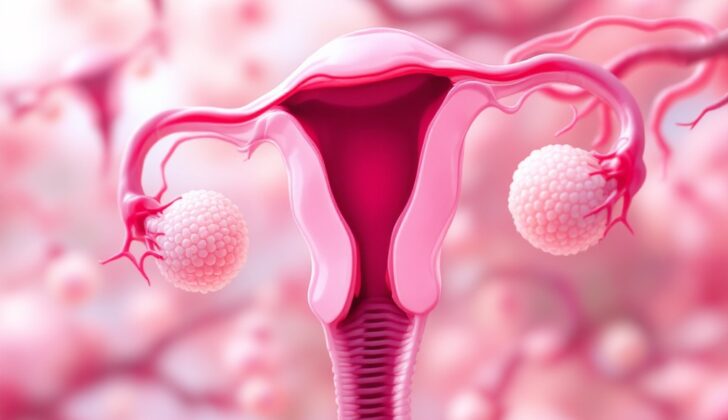
What is Ovarian Cystadenoma?
Epithelial ovarian tumors make up 60% of all ovarian tumors and 40% of non-cancerous ones. These tumors can be harmless (benign), a little risky (borderline), or harmful (malignant). Ovarian cystadenomas are a common type of non-cancerous epithelial tumor which generally have a good outcome. The two main types of this tumor are serous and mucinous cystadenomas, while endometrioid and clear cell cystadenomas are not common.
With all the advancements in scanning techniques, a concrete diagnosis of cystadenomas is mainly done by studying tissue under a microscope from the removed surgical specimen. This easy-to-understand review will specifically discuss ovarian cystadenomas and their characteristics when studied under a microscope.
What Causes Ovarian Cystadenoma?
Serous cystadenomas are a type of growth that don’t have genetic mutations in the KRAS or BRAF genes, unlike some other types of tumors. These growths usually have multiple origins (polyclonal), but can also have a single origin (monoclonal). These tumors begin as an overgrowth from the lining of your organs (epithelial inclusions). In some instances, the tumor cells can show changes in the number of copies of their DNA.
Mucinous cystadenomas, on the other hand, are linked with dermoid cysts. This connection suggests that some of these cysts may start in germ cells, which are the cells in your body that can develop into sperm or eggs. Other mucinous cystadenomas are linked to Brenner tumors, which suggests they could start in the surface lining of your organs. In many of these cysts, there are genetic mutations in the KRAS gene.
Endometrioid cystadenomas are a third type of cyst. Research has shown that these cysts, as well as clear cell and seromucinous tumors, might start in areas suffering from endometriosis, which is a condition where the tissue that normally lines the inside of your uterus grows outside it.
Lastly, according to some experts, seromucinous cystadenomas are believed to develop from endometriosis as well.
Risk Factors and Frequency for Ovarian Cystadenoma
- Serous cystadenoma is a benign tumor of the ovary. It accounts for 16% of all ovarian tumors and two-thirds of benign ovarian tumors. These tumors can affect adults of any age, but they usually develop in people aged 40 to 60. They can impact both ovaries in 10 to 20% of cases.
- Mucinous cystadenoma is a type of benign ovarian tumor. It makes up 80% of mucinous ovarian tumors. These tumors typically develop in women during their thirties to sixties, but they can also occur in younger women. They usually only affect one ovary, with this being the case in 95% of instances.
- Endometrioid cystadenoma is a type of tumor that accounts for 2 to 4% of all ovarian tumors. These tumors typically occur in women in their forties and fifties and often associate with endometriosis.
- Clear cell cystadenoma is a very rare type of ovarian tumor.
- Seromucinous cystadenoma often occurs in adults, usually in the later stages of their reproductive years.
Signs and Symptoms of Ovarian Cystadenoma
Ovarian cystadenomas are types of cysts that can form on the ovaries. These cysts usually range in size from 1 to 3 cm and are often found incidentally, meaning they are discovered during an ultrasound for a different gynecological issue.
When these cysts grow larger, they can cause some nonspecific symptoms such as:
- Pelvic pain
- Bloating
- General discomfort
Testing for Ovarian Cystadenoma
The CA-125 blood test can play an essential role in distinguishing between non-cancerous and cancerous ovarian growths. If the test results, imaging results, and physical examination appear normal, it’s unlikely that ovarian cancer is present.
Several diagnostic imaging methods can help identify ovarian cystadenomas, or cysts. These include pelvic ultrasound, computed tomography (CT scan), and magnetic resonance imaging (MRI).
Certain features visible on these imaging tests can indicate that an ovarian growth is likely benign (noncancerous). These features include cysts that have a single compartment, few dividers, thin walls, and no small, finger-like growths. Unfortunately, the specific cell type of cystadenomas cannot be determined just from these imaging results.
Specifically, pelvic ultrasound is usually the first imaging test used when an abnormal growth is found in the ovaries. Both the transabdominal ultrasound (performed outside the body) and the endovaginal ultrasound (performed inside the vagina) are good options for examining these growths.
CT scans can also be beneficial in diagnosing abnormal growths in the ovaries but may not provide all the necessary information in some cases.
MRIs, on the other hand, can give more detail. Benign (noncancerous) growths in the ovaries are mainly filled with fluid, while malignant (cancerous) growths typically have both solid and fluid-filled areas. In an MRI, fluid-filled areas will appear very bright on the images.
Lastly, to confirm a diagnosis of ovarian cystadenomas, the growth needs to be surgically removed and examined under a microscope, a process known as histopathology.
Treatment Options for Ovarian Cystadenoma
The treatment plan for ovarian cystadenomas, which are fluid-filled sacs that develop on the ovaries, depends on several factors. These include symptoms you’re experiencing, the size of the cyst, your age, your medical history, and whether or not you’ve gone through menopause.
The common medical procedure for treating ovarian cystadenomas is unilateral salpingo-oophorectomy or ovarian cystectomy. A unilateral salpingo-oophorectomy is a surgery to remove one ovary and one fallopian tube, while an ovarian cystectomy is a surgery to remove the cyst from the ovary.
It’s unusual for these cysts to come back after treatment. If they do, it’s likely due either to not all of the cyst being removed during the initial surgery or a new cyst forming altogether.
What else can Ovarian Cystadenoma be?
For diagnosing different types of cystadenomas, doctors need to consider other conditions that might actually be causing the symptoms. These conditions can vary depending on the type of cystadenoma.
For a Serous Cystadenoma, doctors consider these possibilities:
- Broad ligament cysts
- Hydatid cyst of Morgani
- Mesonephric cysts
- Mesothelial cysts
- Extensive cortical inclusion cysts
- Cystically dilated follicles
- Paratubal cysts
- Hydrosalpinx
- Cystic struma ovarii
- Rete cystadenomas
- Polycystic ovarian disease
For a Mucinous Cystadenoma, the doctors look to rule out:
- Cystic mature teratoma
While diagnosing an Endometrioid Cystadenoma, these conditions are considered as an alternative:
- Serous cystadenofibroma
And for Seromucinous Cystadenoma, the differential diagnosis includes:
- Serous cystadenoma
- Mucinous cystadenoma
- Cystic mature teratoma
What to expect with Ovarian Cystadenoma
Serous cystadenomas are essentially harmless lumps or lesions, but after removal through an operation called cystectomy, they might occasionally come back.
Mucinous cystadenomas are also benign or non-dangerous. However, after being treated and removed through cystectomy, there might be cases of them reappearing again.
Endometrioid cystadenomas are harmless lumps which usually result in excellent health outcomes. There’s a slight chance of these tumors reappearing, but it’s quite rare.
Clear cell cystadenoma, which is a benign or non-dangerous cyst, also has an excellent prognosis, meaning that the eventual health outcome is very favorable.
Possible Complications When Diagnosed with Ovarian Cystadenoma
Cystadenomas of the ovary are non-cancerous growths that hardly ever come back even if they are not entirely removed.
Rare issues that can occur with ovarian cystadenomas are:
- Twisting of the ovary
- Bursting of the cyst
If a mucinous cystadenoma, a specific type of cyst filled with a gelatinous substance, bursts, there is a risk of pseudomyxoma peritonei. This condition causes a buildup of mucus in the abdomen.


