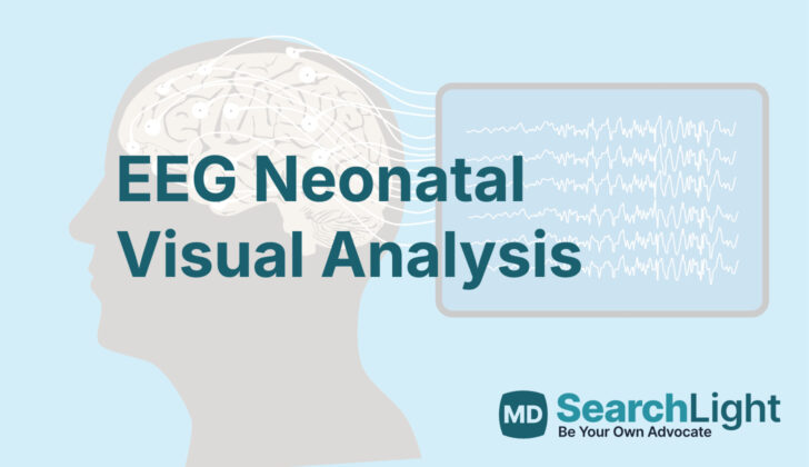
Overview of EEG Neonatal Visual Analysis
An electroencephalogram (EEG) is a test that records the electrical activity of the brain. This test uses small, sticky sensors called electrodes, that are placed on the scalp. Typically, 21 of these electrodes are used to record activity, and one extra is used as a reference point. These electrodes are placed in specific places on the head and are marked by odd numbers for the left side, even numbers for the right side. Letters represent different areas of the brain (F for frontal, C for central, T for temporal, O for occipital, and Z for midline).
In newborn babies, an EEG can give important information about how the brain is developing. It can also help identify if there are signs of overly active brain activity, abnormal electric patterns, or seizures. Interpreting the EEG for babies can be quite tricky, requiring more skills and training, as there are normal variations at different stages of development that can be confused with abnormal patterns. For a baby’s EEG, the arrangement is modified to use only eight electrodes and a slower recording speed. Analyzing the visual pattern of the recorded brainwaves is considered the best method for interpreting EEG results in newborns.
It’s important to remember that every individual’s brain activity can look a bit different, so understanding the results often requires careful visual analysis. Even though there are software programs that can identify brainwave patterns, the human interpretation is an essential part of the process. This visual analysis provides a comprehensive overview of the main features of the brain’s electrical activity to draw conclusions and make clinical correlations. This piece explains these visual analysis techniques as they apply to newborn babies.
Anatomy and Physiology of EEG Neonatal Visual Analysis
The brain creates electrical activity that are caused by different areas of the brain communicating with each other. These messages can either be excitatory, meaning they encourage more brain activity, or inhibitory, which means they reduce brain activity. These messages are mostly created by cells called pyramidal cells. We can record these messages and see a pattern of activity between two brain areas over time. A rhythm is then created in these patterns, which can be seen on an EEG, which is a machine that measures brain activity.
Newborns spend most of their time sleeping, so it’s important to study what their brain activity looks like during this time. Sleep has two types – active sleep, which is similar to REM (rapid eye movement) sleep in adults and is when most dreaming happens, and quiet sleep, which is similar to non-REM sleep in adults, which is a deeper, more restful form of sleep. There have been some interesting findings in the brain activity of newborns in the first 6 hours of their life compared to 48-72 hours after birth. These findings include changes in their sleep-wake patterns, appearance of sporadic sharp waves in brain activity, and an overall increase in the orderly flow of brain activity over time. This suggests the newborns’ brains are adjusting to life outside the womb. A similar pattern is seen in babies that are born with a lower than average birth weight compared to babies with a normal birth weight.
Why do People Need EEG Neonatal Visual Analysis
If a baby is showing unusual movements of the face or limbs, issues with muscle tone, difficulty with feeding, unusually low scores on newborn health checks (known as Apgar scores), changed awareness levels, or strange results from brain scans, a test known as an electroencephalogram (EEG) might be required. This test is often used when health professionals think the baby might be experiencing seizures. An EEG is a useful tool for checking on a baby’s brain development, spotting overactive brain areas, identifying seizure activity, or predicting future health outcomes.
An EEG provides a way for doctors to monitor the growth and function of a newborn’s brain as they grow each week. In order to properly interpret the EEG results, doctors need to take into account several factors. These include the baby’s gestational age (the time the baby spent growing in the womb) plus the number of weeks since the baby was born (this is called the postconceptional age), the shape and thickness of the baby’s skull and scalp, the resistance between the EEG sensors, any normal variations for the baby’s age, any external factors that might interfere with the EEG readings, and the baby’s sleep patterns.
Note that full term pregnancies usually last between 38 to 42 weeks. If a baby is born before completing 38 weeks of gestation, this is considered a premature birth.
When a Person Should Avoid EEG Neonatal Visual Analysis
The only people who should not have this procedure are those who have issues with their skin which stop the electrodes from being able to stick to their head. This might be because of cuts, scrapes, or other problems with their skin.
Equipment used for EEG Neonatal Visual Analysis
Recording brain waves, or EEGs, are slightly different for babies compared to adults. This is mainly due to the baby’s smaller head size and certain areas of the brain not being as active. Special equipment is needed which includes eight sensors (called electrodes) that are placed on the scalp, a heart monitor (EKG), a muscle activity detector (EMG), monitors for eye movements, and breathing monitors. Additional sensors can be used if needed to cover the middle of the head. This setup allows us to effectively monitor the brain regions where most activity occurs in babies.
Who is needed to perform EEG Neonatal Visual Analysis?
Having skillful EEG technicians is crucial for making sure that the electrodes, which help measure brain activity, are correctly placed and the results recorded accurately. These technicians should know how to recognize and correct issues that could affect the accuracy of the results, like misplaced electrodes. They aim to reduce any factors that could cause misleading activity. In addition to the EEG technicians, care for this process involves a team of different specialists. This includes nurses who specialize in caring for newborns in critical health conditions, primary care providers, consultants, as well as respiratory therapists who make sure the patient’s breathing is maintained. This team works together to deliver the most accurate results from the continuous video EEG, which monitors ongoing brain activity.
Preparing for EEG Neonatal Visual Analysis
When recording an EEG (a brain scan) on a newborn baby, here are some key steps:
– Limit any movement or excitement for the baby. This is important as it allows the EEG to capture an accurate reading of the baby’s brain activity.
– Make sure you have all the right equipment, such as electrodes and other important tools, ready and set up.
– Chart down important information like the baby’s head positioning, age since conception, other health conditions, and more. This information is important to help interpret the EEG results.
– Try to reduce any disturbances or ‘artifacts’ that may cause issues in the EEG recording.
– Apply the EEG electrodes properly on the baby’s skin so that they don’t damage the skin and ensure the electrical resistance, also known as ‘impedance’, is less than 10,000 ‘ohms’. The electrodes are used to detect and record brain waves. It’s important to make sure they’re attached securely and comfortably.
How is EEG Neonatal Visual Analysis performed
When a newborn baby’s brain activity is being recorded in a process called an EEG, less electrodes are used than in adults because of the baby’s small head size. The electrodes are placed on the baby’s head in different areas: forehead to side of the head, forehead to the upper middle part of the head, side to back of the head, and middle of the head to back of the head. The space between the electrodes on a baby’s head is usually around 40% of the head’s diameter. The machine also records muscle movements (EMG), heart activity (EKG), and breathing, to help tell the difference between any unusual signs (epileptiform activity) and normal body functions.
The electrodes for the eyes are placed just 0.5 cm from the outer corner of the eyes. Certain settings on the EEG machine need to be adjusted to make sure that anything that isn’t brain activity- called artifact- can be reduced or eliminated. This involves changing the recording speed (it’s usually slower than for adults), using filters and setting amplifier sensitivities. These settings for the EEG, EMG, and eye electrodes can be changed as necessary. Everything is done to accurately capture the unusual sharp spike and slow waves that interrupt the regular background brain activity. A recording period of 2 to 3 hours is recommended, so that brain activity can be tracked during periods of wakefulness and sleep.
Some factors can lower the voltage of the brain activity recorded in the EEG. These include cooling therapy which can be applied to either the head or the whole body, and some medicines including sedatives, epilepsy drugs, and morphine. The pattern of eye movements and breathing helps determine the different sleep stages the baby is going through. Lastly, various disturbances like the heart’s activity (EKG), pulse rate, nursing care, caregiver activity, breathings, ventilator status, incubator noise, physical movements, loud sounds, and feeding, can cause irregularities in the EEG recording.
Possible Complications of EEG Neonatal Visual Analysis
Problems can often occur during long-term health monitoring and these can include:
* Breakdown of the skin
* Skin infections
To protect the skin and prevent infections, the use of things like moisturizers, gels, and topical antibiotics (medications applied to the skin) can be effective.
What Else Should I Know About EEG Neonatal Visual Analysis?
Newborn seizures are fits or convulsions that can occur from the time a baby is born up to about 28 days of age. This time is the most likely for a baby to have seizures, with 80% happening within the first 24 to 48 hours of life. It’s important to account for the baby’s intended due date when monitoring their development, since some newborns may be born earlier or later than expected.
Seizures in newborns might be a sign of lasting brain damage, so it’s important to detect and treat them as soon as possible. Around 3 in every 1,000 full-term newborns, and up to 132 in every 1,000 premature babies, will have seizures within their first two days of life. Seizures in newborns can have different symptoms and severity, which helps doctors classify and treat each case properly.
Often, subtle seizures can go unnoticed, with symptoms like unusual eye movements, repetitive mouth movements, difficulty breathing, and sudden tongue thrusting. Other types of seizures can involve rhythmical muscle contractions (clonic), sudden spasms of the limbs (myoclonic), sustained muscle stiffening (tonic), or alternating muscle stiffening and contractions (generalized tonic-clonic).
Newborn’s brain activity can be monitored using a test called an EEG, which looks at their brain waves. Brain waves can change depending on whether the baby is awake, quietly asleep, or in a deeper sleep stage, and can provide doctors with important information about the baby’s neurological development.
For example, when a baby is awake, their eyes are open with rapid movements, irregular and active respiration, rapid startles, and large body movements. When the baby is in quiet sleep, they have closed eyes, lack of eye movements, no muscle tension, and regular respiration. During active sleep, the baby may show rapid eye movement, occasional frowns or sucking behavior while asleep.
EEG patterns can look different depending on the baby’s gestational and corrected age. At different stages, the baby’s brain waves will exhibit different frequencies, amplitudes, and patterns of activity. This data helps doctors to understand if the baby’s brain development is on track for their age.


