What is Compressive Optic Neuropathy?
Compressive optic neuropathy, or CON, is a condition where any pressure along the optic nerve, whether internal or external, affects the nerve. This condition can also result from various other factors such as lack of oxygen, metabolic issues, or physical damage. The most common symptom is a gradual loss of sight in one eye, sometimes accompanied by headaches, while both eyes may be affected by certain central brain or bilateral orbital pathologies. It’s very important to correctly identify the cause of CON because many different diagnoses may apply, each requiring a different treatment strategy.
The optic nerve is incredibly complex with over a million nerve fibers. This complexity reflects the crucial role vision plays in our evolution. Our visual processing system begins at the retina, the light-sensitive layer at the back of the eye, and ends in the visual cortex at the back of the brain. The retina itself is comprised of it visual and non-visual parts, with the visual part consisting of neural and pigmented layers, while the non-visual retina extends from the pigmented layer and terminates in the areas related to the color and shape of the eye.
The optic nerve starts in a part of the sclera, or the white part of the eye, and leaves the eye socket through a passageway known as the optic canal. When it exits the socket, the optic nerve is covered by layers that make up the optic nerve sheath. Axons running from the ganglion cells, a type of neuron, form the optic nerve and cross over at the optic chiasm. They then follow a path to the lateral geniculate ganglion, a relay center in the brain, continuing onwards to the primary visual cortex. This journey is approximately 50mm long, with segments within the eye, inside the eye socket, within the optic canal, and inside the brain.
What Causes Compressive Optic Neuropathy?
Compression of the optic nerve (CON) can happen because of obstructions either inside or outside the nerve. In rare cases, a growth within the nerve, such as optic nerve glioma, can slowly compress and damage its fibers. There can be many causes of optic nerve compression, and they are typically categorized as follows:
1. Infectious causes can include something like Aspergilloma, which is a type of fungal ball.
2. Inflammation-based causes can include conditions like idiopathic orbital inflammation (also known as a pseudotumor), Sarcoidosis, and Graves’ disease (also known as thyroid orbitopathy).
3. Vascular conditions that can cause optic nerve compression include an aneurysm, carotid-cavernous fistula, lymphangioma, orbital varix, and an enlarged internal carotid artery.
4. Trauma-related causes can include a hematoma (blood clot) or fracture.
5. Tumor-related causes include a range of tumors such as hemangioma, schwannoma, meningioma, glioma (common in Neurofibromatosis Type 1), lymphoma, sarcoma, metastatic tumors, pituitary adenoma, craniopharyngioma, and dermoid.
6. Bone tumors or lesions can also occur and these include fibrous dysplasia, Paget’s disease, osteoma, osteopetrosis, hyperostosis, and Langerhans cell histiocytosis.
7. Other causes include a mucocele (a swelling filled with mucus) and granulomatous meningitis (a type of inflammation in the membranes of the brain and spinal cord).
Risk Factors and Frequency for Compressive Optic Neuropathy
Statistics show that there are roughly 4 cases of CON (a specific type of disease) per 100,000 people every year. However, these rates can change depending on a person’s age, sex, race, and ethnicity. Also, it’s worth noting that these rates can also vary depending on the cause of the disease.
- Thyroid orbitopathy, a complication of Graves’ disease, affects 16 out of 100,000 women and about 3 out of 100,000 men.
- Craniopharyngioma, a type of brain tumor, occurs in 0.5 to 2 out of every 100,000 people each year.
- Fibrous dysplasia, a bone disorder, affects 1 in 5,000 to 10,000 people.
- Orbital hemangiomas, benign tumors around the eye, are found in 8 to 10 percent of children’s benign tumors and are five times more common in girls than boys.
- Pituitary adenomas, a type of brain tumor, have a 10.5% occurrence rate but only cause vision problems when they grow large enough to press on the optic nerve.
- Optic gliomas, another type of brain tumor, commonly occur in children and makeup about 3 to 5 percent of childhood tumors.
- Cerebral aneurysms, a bulging blood vessel in the brain, are found near the optic nerve in about 3 percent of the cases.
Signs and Symptoms of Compressive Optic Neuropathy
People with CON (chronic optic neuropathy) usually experience a gradual loss of vision, which can affect one or both eyes. Additional symptoms can include headaches, nausea, vomiting, double vision, color perception issues, eye bulging, unusual pupil reaction to light, sensitivity to light, a red eye, or unexplained weight loss. In rare cases, patients may experience sudden or rapid loss of vision, typically arising from incidents like blunt force trauma or penetrating injuries to the eye area. The areas most susceptible to injury are the orbital apex and the optic canal.
It’s crucial for doctors to get detailed information about a patient’s medical history and conduct a thorough physical exam to help pinpoint the cause of their symptoms.
- Details of vision loss: Is it the same in both eyes or different?
- The onset of vision loss: Did it start slowly or quickly?
- Family history of cancer
- Prior exposure to radiation
- Risk factors for heart disease: high blood pressure, peripheral vascular disease, tobacco use
- Any underlying metabolic diseases
- History of autoimmune diseases
The physical examination usually includes:
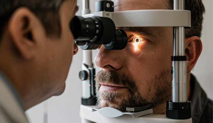
- Snellen chart: a test for visual sharpness
- Funduscopic exam: an examination of the back of the eye
- Slit-lamp examination: an assessment of the retina, retinal arteries and veins, cornea, fovea and optic cup
- Visual field test: a test to check central vs. peripheral visual loss
- Ishihara’s test: an assessment to identify color vision issues
- Tonometry: a test for eye pressure
- A check for eye movement
- Assessment of eye protrusion
Testing for Compressive Optic Neuropathy
Doctors should perform a thorough assessment of the nervous system followed by a detailed evaluation of the eyes. The purpose of the examination is to establish initial benchmarks for how well one can see. This will enable doctors to monitor any changes in vision.
The eye affected by optic nerve compression might exhibit decreased vision and potential issues with color perception, a condition called dyschromatopsia. Color perception can be tested using the Ishihara test plate, a well-known method for detecting color blindness. If the eye bulges out or resists pressure when touch manually, this may suggest the presence of a lesion inside the eye socket. An examination of how the eyes move can also reveal abnormalities. While the optic disc (the part of the eye connecting to the optic nerve) can exhibit signs of shrinkage or swelling, in some cases it may appear normal. Sometimes, abnormal blood vessels may appear in the eye due to blockage of the venous return.
Several laboratory tests may be ordered, including full blood count, comprehensive metabolic panel, lipid profile, thyroid tests, hormone tests and markers for bone metabolism and prostate health. For instance, a higher level of angiotensin-converting-enzyme is detected in over half of the patients with active sarcoidosis, a disease affecting numerous body parts.
Magnetic resonance imaging (MRI) of the brain and eye sockets can provide a detailed view of the optic nerves and surrounding structures. Specific images are required to display lesions inside the eye sockets. A computerized tomography (CT) scan of the head and eye socket is recommended if lesions involve the bones around the eye. This is also useful for viewing fractures and associated injuries following trauma. For lesions located at the front of the eye socket, an ultrasound can be used to obtain a biopsy, a small sample of tissue for testing.
Treatment Options for Compressive Optic Neuropathy
The first part of treating a condition involves dealing with the core issue itself. Corticosteroids, a type of medication, can be used to reduce inflammation for conditions like sarcoidosis and thyroid disease. However, stopping the use of these steroids suddenly can lead to a rapid worsening of vision. In some cases, an operation known as orbital decompression can help improve thyroid-related vision problems.
For certain tumors closely attached to the optic nerve, such as optic nerve meningiomas, surgery might worsen vision loss. On the other hand, radiation therapy may be useful for aggressive, recurring tumors, especially those near critical brain and cranial nerves. It can also be used for tumors that are hard to reach with surgery, like those in the cavernous sinus. But care must be taken as radiation can cause irreversible damage to the optic nerve.
In the case of traumatic injury, mild cases often improve on their own, so a wait-and-see approach is usually recommended. Steroids haven’t shown to be beneficial in these cases. Surgery may be performed if there’s evidence of compression on medical imaging. If the optic nerve is directly compressed by fragments of bone or a pocket of blood under the outer layer of the bone (subperiosteal hematoma), surgery is typically the chosen treatment. However, surgery may come with risks, including complications like leakage of cerebrospinal fluid (the fluid around the brain and spinal cord) and meningitis (a potentially serious infection).
What else can Compressive Optic Neuropathy be?
When looking for the specific cause of certain eye conditions, doctors need to consider a variety of potential factors. These might include:
- Glaucoma
- Ischemic optic neuropathy, which relates to the blood flow to your eyes
- Retinal vein occlusion, a blockage of blood flow in your eyes
- Multiple Sclerosis
- Uveitis, inflammation of the middle section of your eye
These disorders need to be examined because the right treatment can vary depending on the specific cause.
Moreover, it is also important for the doctor to eliminate the following possibilities, as each of these conditions requires different management:
- Pituitary tumors
- Schwannoma, a tumor of the tissue that covers your nerves
- Meningioma, a tumor that arises from the membranes surrounding your brain and spinal cord
- Aneurysm, an enlarged blood vessel
- Arteriovenous malformation, a tangle of abnormal blood vessels
- Lymphoma, a type of blood cancer
- Sarcomas, a type of cancer that starts in bone or muscle
- Metastasis, where cancer has spread from one part of your body to another
- Glioma, a type of brain or spine tumor
- Injury to the brain or spine
- Sarcoidosis, a condition where small collections of inflammatory cells grow in the body
What to expect with Compressive Optic Neuropathy
The likelihood of improvement in CON, a form of eye disease, hinges on its root cause (whether it’s due to poor blood supply or nerve damage) and how quickly treatment begins.
The speed of easing of vision-related symptoms generally lines up with how soon the eye is relieved from any pressure it’s under. Most agree that sooner is better when it comes to this relief. In some cases, full recovery of vision can occur as early as one week. And even a slow but steady improvement in vision has been observed. Moreover, over half of the patients seem to get better, regardless of how bad their vision was before the treatment. As such, it’s advised to start the pressure relief procedure early, but positive outcomes have also been seen when it’s performed later on.
It’s key to note that the prospects for vision recovery can vary greatly, depending on the specific reason causing the compression on the optic nerve. When it’s due to a tumor, factors that can hinder vision improvement include the severity of vision loss, disc atrophy, having to remove the tumor multiple times, the tumor extending to the cavernous sinus, a hard tumor, the absence of an arachnoid plane, extensive removal of the tumor, and a long period of visual loss. Nevertheless, about 60% of patients with tumors will see some level of improvement after the pressure on the optic nerve is relieved.
Possible Complications When Diagnosed with Compressive Optic Neuropathy
CON, also known as Chronic Open-Angle Glaucoma, can lead to a series of complications. These can include swelling of the optic disc (papilledema), blurred vision, issues with the pupils’ response to light (afferent pupillary defect), double vision (diplopia), nausea, vomiting, permanent vision loss, difficulty moving the eyes in different directions (impaired extraocular movement), and challenges in daily activities due to vision problems.
Common Complications of CON:
- Swelling of the optic disc (papilledema)
- Blurred vision
- Issues with pupils’ response to light (afferent pupillary defect)
- Double vision (diplopia)
- Nausea
- Vomiting
- Permanent vision loss
- Difficulty moving the eyes in all directions (impaired extraocular movement)
- Challenges in daily activities due to vision problems
Decompressive surgery, a treatment for this condition, can also have serious complications like bleeding (hemorrhage), infections, total loss of vision, having a breathing tube for too long (prolonged intubation), unconsciousness (coma), and in worst cases, even death.
Potential Complications of Decompressive Surgery:
- Bleeding (hemorrhage)
- Infections
- Total loss of vision
- Having a breathing tube for too long (prolonged intubation)
- Unconsciousness (coma)
- Death
Preventing Compressive Optic Neuropathy
If a patient’s vision is severely impacted, they should be advised to wear protective glasses to safeguard the eye that isn’t affected. Regular check-ups with an eye doctor are crucial for all patients who have CON (compressive optic neuropathy) to monitor any potential improvements or declines in vision.
Sufferers of CON will need thorough checks from a team of healthcare professionals. This is due to the range of factors that can cause CON, and to ensure that any necessary treatments are administered as quickly as possible to improve the patient’s condition.
Early surgery to relieve pressure (decompression) is normally suggested, but satisfactory results can still be achieved even if the operation is delayed. Patients should arrange an appointment with an eye doctor as soon as they notice any symptoms. In over half of the cases, patients have reported an improvement, regardless of how their vision was before surgery.
For those with tumors, 60% of patients have noted some improvement in their condition after surgery to relieve pressure on the optic nerve.


