Overview of Care of a Chest Tube
The treatment and care for chest tubes, also known as thoracostomy tubes, are directed and managed by the doctor who’s taking care of the patient. So, it’s pretty tough to provide a single set of instructions that could fit everyone’s situations. It is a good idea for a patient to have a detailed discussion about what to expect and how the tube will be managed with their doctor. There might be specific guidelines at the hospital or clinic that could provide more information on how this treatment is handled. This discussion is about how chest tubes are placed surgically and how they are taken care of after the procedure.
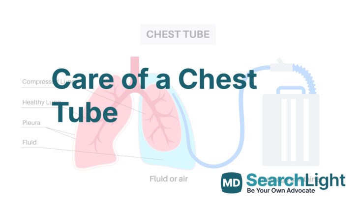
Anatomy and Physiology of Care of a Chest Tube
The thorax, or chest area, contains all the organs surrounded by the ribs and chest bones. It’s separated from the stomach area by a muscle called the diaphragm. Inside the chest, there are two spaces known as the pleural spaces, with one on the left and one on the right, divided by a central region called the mediastinum. Each of these spaces holds a lung – the left lung has two sections (lobes), and the right lung has three.
When we breathe in, a downward movement of the diaphragm creates some negative pressure, or a kind of vacuum, inside the lungs. This causes air to rush into the lungs to balance everything out. Breathing out, in contrast, is usually just a passive process that happens naturally when the chest wall goes back to its original position.
The inner layer of the chest wall is lined with a type of tissue called parietal pleura, and the lungs are lined with another type called visceral pleura. There’s a small amount (about a spoonful to a shot glass) of a special fluid between these layers that helps everything move smoothly when you breathe.
A pneumothorax happens when air gets into the pleural space. As more air is introduced, it takes up space that’s normally filled by the lung, making it so the lung can’t fully inflate with air. If the pressure keeps building, the lung and other tissues in the center of the chest can start to shift over to the other side. In the worst cases, called a tension pneumothorax, this pressure can compress a large vein called the inferior vena cava, which disrupts the return flow of blood to the heart.
Why do People Need Care of a Chest Tube
A thoracostomy tube is a medical device that doctors use for several reasons. In simple terms, it’s a tube that helps remove harmful substances like air or fluids from the area around your lungs. This area is known as the pleural space.
Sometimes, air may fill up in this space, which can prevent the lungs from expanding fully. This is called a pneumothorax. If it gets larger or the doctor is worried it might cause tension (putting dangerous pressure on your lungs and heart), it might be necessary to place a thoracostomy tube. Recent studies have shown that smaller air spaces, less than 35 mm, can also be monitored without needing tube placement. Furthermore, this kind of tube is commonly used after certain heart and lung surgeries.
If the doctor is worried that you have a tension pneumothorax, which is a severe type of pneumothorax, they might need to place a thoracostomy tube right away to relieve the pressure. If placing a tube isn’t possible, they might need to relieve the pressure with a needle or their finger.
On some occasions, the space around the lungs fills up with fluid. This can happen due to a hemothorax, which is blood in the pleural space if the volume is greater than 300 cc. Another potential cause is a pleural effusion, where the fluid could be due to a benign (non-harmful) or a malignant (cancerous) disease. The fluid can put pressure on your lungs, causing symptoms like difficulty breathing.
There are other reasons why a doctor would consider placing a thoracostomy tube. For example, in a condition called chylothorax, where a type of bodily fluid called chyle leaks into the pleural space, a thoracostomy tube along with diet modifications could be part of the treatment plan. Sometimes, an infection in this space, known as empyema, should almost always be treated with surgery and a tube to help drain out the infected fluid. Medical management alone usually doesn’t work in these cases.
Lastly, the tube can be used to deliver medicine or fluid directly into the pleural space. For instance, doctors might use it to warm up the body by circulating warm fluids through this space. They might also use it to bring the lung coverings (the visceral and parietal pleura) closer together, a procedure known as pleurodesis, by putting substances like talc through the tube to cause scarring. Another use might be to deliver chemotherapy drugs directly into the space around the lungs for certain types of cancer like pleural mesothelioma and metastatic disease.
When a Person Should Avoid Care of a Chest Tube
There are no definitive reasons that a person should not have a chest tube placed. However, there are some situations where doctors need to be extra careful about the procedure. One of these situations is when the person has a bleeding disorder caused by blood-thinning medication or a problem with their body’s clotting system, which helps stop bleeding. This makes it harder for their body to stop bleeding, so the risk of the procedure might be greater.
Another situation where extra care is needed is when the patient has had chest or open heart surgery in the past. This is because chest tube placement can cause complications if not done carefully. Additionally, people who have had a procedure called pleurodesis, which involves sealing off the space between the chest wall and the lung to prevent fluid buildup, need extra evaluations to make sure the chest tube can be inserted safely. The chest tube needs to be placed in a part of the chest wall that is not stuck to the lung tissue, to avoid any complications.
Equipment used for Care of a Chest Tube
Before placing a chest tube, different tools and equipment might be needed. This can change depending on the location it’s being done, how urgently it’s needed, the reason it’s being done, and the habits of the healthcare provider. For more details, please see our related articles on chest tubes and chest trauma.
In emergency setups designed for treating trauma, they often have ready-to-use chest tube kits. Other healthcare settings might have different tools or follow different rules. Here are some things to think about before starting the procedure:
A chest tube: Research shows that there’s no significant difference between small chest tubes (20 to 22 Fr size) and the traditionally bigger 28 to 32 Fr tubes from earlier guidelines, as long as they’re used correctly.
A chest drainage system: All modern systems are based on the original 3-bottle system. The chest tube connects to the first bottle, and then other bottles connect one after the other to a suction device. The first bottle collects fluid from the chest; the second bottle acts like a one-way valve stopping air and fluid from going back into the chest, and the third controls suction.
Antibiotics: It’s recommended to take antibiotics beforehand to prevent infections. The kinds commonly used (cefazolin, clindamycin) are designed to prevent infections from bacteria found on the skin. Your healthcare institution will have guidelines on which antibiotics to use. Note that continuing antibiotics after the procedure generally hasn’t been found to reduce infection rates and is usually not recommended.
Other tools needed include: a scalpel for the first cut in the skin (often a #10 blade), various surgical instruments to help with cutting and separating tissue, a large clamp for entering the space around the lungs without cutting (Rochester Peon or Kelly clamps could be used), and something to keep the chest tube attached to the patient.
Who is needed to perform Care of a Chest Tube?
The procedure only needs one person who is permitted and trained to do it. This individual is known as a clinician. If possible, a second person can help out by handling the tools, holding the tube in place while it’s firmly attached, and gently pulling back extra soft or breast tissue. Sometimes, a type of anesthesia called moderate sedation is used to make you feel relaxed and sleepy. If this is the case, a dedicated nurse or another clinician is needed to keep an eye on your airway (the path that air follows to reach your lungs) to make sure you’re breathing properly.
Preparing for Care of a Chest Tube
Prior to inserting an emergency chest tube, it’s important to ensure that the skin is clean and that the patient has received a systemic anti-microbial treatment to prevent infection. If it’s extremely urgent, this cleansing process can be done quickly by pouring iodine over the patient’s skin in the area where the tube will be inserted. To make the patient more comfortable during the procedure, an appropriate pain reliever and sedative are administered. This can be especially helpful when the chest tube insertion is scheduled or done on an urgent basis.
To reduce discomfort even further, a generous amount of local anesthetic can be used. Care is taken to ensure the amount does not exceed safe limits. A common practice is to apply the anesthetic to the spot where the incision will be made and along the pathway where the tube will be inserted, especially on the periosteum, which is the layer covering the rib cage where pain receptors are located.
Before the procedure begins, the person performing the procedure or a member of the support staff should make sure that all the equipment needed for the procedure is in place, working properly, not expired, and there’s backup equipment on standby.
In emergency situations, like in trauma cases, it is generally recommended that all patients should have: an intravenous (IV) line for delivering fluids and medications, supplemental oxygen with monitoring to ensure they are getting enough oxygen, monitoring of blood pressure and other vital signs, and equipment for securing a patient’s airway at the bedside, just in case the patient has difficulty breathing.
How is Care of a Chest Tube performed
A chest tube is usually inserted on the side where there is a problem. The common spot for this is in the area of the fourth or fifth rib (around the nipple line for men and around the line beneath the breast for women). This is usually towards the front and side of your torso. The chest tube helps to relieve pressure or remove fluid from your chest. It might be positioned towards the front of your chest if you have a buildup of air (pneumothorax) or towards the back if you have a buildup of fluid.
To place the chest tube, your doctor will make a small cut in your chest. They will carefully insert the tube through this cut, making sure they avoid any important structures inside your chest such as blood vessels or nerves. They might use a special tool to help open up a pathway for the tube. You might feel a bit of discomfort when the tube is inserted, but your doctor will do everything they can to reduce your pain.
Next, the doctor will insert a finger into the incision to make sure the tube can fit, and to break up any blockages that might prevent the tube from working properly. The doctor will then measure how far the tube needs to go into your chest so that it can do its job effectively. Once the tube has been inserted to the right depth, it will need to be secured to your chest. This could be done with stitches, tape, or a special appliance.
The tube is then attached to a chest drain system, which might be connected to suction to help remove the air or fluid from your chest. The success of the treatment can be gauges by measuring the amount of fluid that is removed. If a lot of fluid is rapidly removed, it may suggest a serious injury that needs to be surgically repaired. After the tube is inserted, an x-ray will be taken to confirm that the tube is in the right place inside your chest.
If fluid build up in your chest doesn’t improve, the doctor might decide to use a kind of treatment that dissolves blood clots (thrombolytic therapy). This treatment can help to break down blood clots or thickened fluid to help it drain out more easily. This treatment has been linked with a reduced need for surgery but hasn’t been shown to help people live longer. As of now, there isn’t a standard protocol for using this treatment, and it is still being studied to find the best way to use it.
After the tube is placed, your medical team will monitor you. They’ll listen to your breathing, check the volume of fluid being collected by the tube, and look at the tubing and the collection chamber. You might also get daily x-rays if you have a build up of air in your chest (pneumothorax). They might also use a special type of ultrasound to assess your chest if you continue to have a buildup of air in your chest. If the fluid in the tubing seems to be moving in time with your breaths, it suggests that the tube is working properly.
Throughout your treatment, your medical team will look for any leaks in the tube or tubing, or signs of a leak within your chest. Most leaks will seal by themselves over time and can be tracked by seeing less and less bubbling over the days. For larger leaks that don’t seal by themselves, surgery might be needed.
The chest tube may need to be adjusted or cleared of any potential blockages from time to time. This might involve gently massaging the tube to create suction and pull any blockages into the collection chamber. This is done by holding the tube near the incision site to prevent it from being pulled out, and then moving down the length of the tube while squeezing it. Some doctors might request this technique, although it doesn’t have strong evidence supporting its use.
Possible Complications of Care of a Chest Tube
Putting a chest tube can sometimes cause serious issues, some of which can even be life-threatening. Doctors are cautious about potential injury to any organs or structures near where the tube is placed. These organs include lungs, diaphragm (the breathing muscle at the bottom of the chest), esophagus (the food pipe), heart, liver, and spleen.
The diaphragm is quite sensitive and can easily be injured, and this can lead to the chest tube entering the space with your stomach organs. Also, it’s important to remember that during the procedure, blood vessels could be accidentally injured, which might result in slow or severe bleeding within the chest region. The blood vessels at risk include large vessels near the heart, arteries on the side of the chest, arteries near the shoulder, blood vessels between the ribs, blood vessels inside the chest wall, and blood vessels in the lungs.
On top of this, if the problem that was taking up space in the chest isn’t getting better, it might mean that the tube is outside the space it’s supposed to be in within the chest or that a sealed-off pocket within the chest wasn’t properly relieved of pressure.
What Else Should I Know About Care of a Chest Tube?
If conditions that occupy space in the chest area, like tumors or cysts, aren’t treated, they can eventually cause a serious medical condition where the pressure in your chest builds up. This can lead to an emergency situation where your heart or lungs stop working because they’re not getting enough blood coming in (decreased preload). This is why it’s so important to identify and treat these conditions early.


