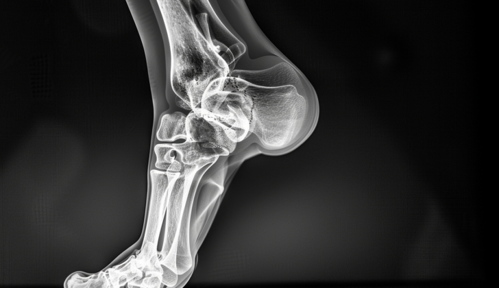
What is Bimalleolar Ankle Fractures?
Bimalleolar fractures refer to a particular kind of ankle fracture that affects both sides of your ankle. These areas are the ends of two bones known as the fibula (on the outside of the ankle) and the tibia (on the inside). These two bones connect with another bone called the talus, forming what we often call the ankle joint. This joint is kept stable by ligaments, or strong bands of tissue, on both sides, helping maintain alignment and keep the fibula and tibia together. Bimalleolar fractures may also damage these ligaments. Even if only the ligaments are affected rather than a break in either of the bones, it still results in an unstable fracture that may need to be treated with surgery.
What Causes Bimalleolar Ankle Fractures?
Based on the Lauge Hansen classification system, the most common cause of bimalleolar fractures, which are fractures involving two bones in the ankle, is from supination and external rotation injuries. Essentially, this means the ankle gets turned upwards and twisted outwards. In addition, eversion, or the act of turning the sole of the foot outward, is recognized as the most common action responsible for all the damage in these types of fractures.
Risk Factors and Frequency for Bimalleolar Ankle Fractures
Ankle fractures are relatively common, making up 9% of all bone breaks. In the United States, they’re the most common type of lower limb fracture and are very frequently seen in emergency rooms. The bimalleolar fracture, which is a specific type of ankle fracture, accounts for 60% of these incidents, affecting 187 out of every 100,000 people. This type of fracture is most common in older women and young men. When we look at all fractures, ankle fractures are the third most common overall, and among athletes, they are the most common. Also, in patients over 60, ankle fractures are the third most common type of fracture.
- Ankle fractures make up 9% of all fractures.
- In the US, they are the most common type of fracture in the lower limbs.
- They are very frequent in the emergency room.
- The bimalleolar fracture is a type of ankle fracture making up 60% of all ankle fractures.
- This fracture affects 187 out of every 100,000 people.
- It is most often seen in older women and young men.
- When considering all kinds of fractures, ankle fractures are the third most common.
- They are most common among athletes.
- In patients over 60, ankle fractures are the third most frequent fracture.
Signs and Symptoms of Bimalleolar Ankle Fractures
When visiting a doctor with a suspected injury, they will first talk with you about your medical history to identify any existing conditions that could interfere with healing. They will be particularly interested in conditions such as diabetes, poor blood circulation, weak bones, alcohol consumption, smoking, or cancer. They will also be interested in any medications you may be taking that could slow the healing process, like corticosteroids. The doctor will also want to know details about the injury itself, like when and how it happened.
During the physical examination, the doctor will examine your wounded limb looking for any obvious abnormalities or changes in the skin that might show a change in blood flow or indicate a wound that might suggest a broken bone. They will also check your knee, fibula, tibia, ankle and foot for signs of a fracture such as swelling, redness, hematoma (blood pooling beneath the skin), and tenderness. They will see if you can bear weight on your injured foot, and use touch to identify the fracture’s exact location. Furthermore, to ensure the blood flow and nerve function in your foot and ankle haven’t been affected, they will check your foot’s pulse, motor skills, feeling, and capillary refill time (how quickly your skin returns to its normal color after being pressed).
The anatomy of the ankle includes the tibia, fibula, and talus bones connected and supported by strong ligaments. On the inner (medial) side of the ankle, the deltoid ligament is important. It is triangular in shape and made up of four smaller ligaments that stabilize the medial side of the ankle by anchoring the tibia to the foot. If this ligament ruptures in an ankle fracture, it can cause joint instability. On the outer (lateral) side of the ankle, there are three important ligaments: the anterior talofibular ligament, calcaneofibular ligament, and posterior talofibular ligament. These ligaments anchor the fibula (the smaller, outer bone of the lower leg) to the foot and help prevent the ankle from twisting inwards too much.
The syndesmosis is another significant part of the ankle’s anatomy. It’s a long membrane that ties together the two lower leg bones (the tibia and fibula). The syndesmosis plays a crucial role in keeping the ankle joint stable and can be damaged in certain types of ankle injuries, particularly those involving excessive upward bending of the foot or rotation of the ankle.
Testing for Bimalleolar Ankle Fractures
According to the Ottawa Ankle Rules, a doctor shouldn’t ask for ankle X-rays unless there’s pain or tenderness in the ankle bones coupled with either sensitivity at the tip of the ankle bones or an inability to put weight on the foot after being injured.
X-ray imaging is considered the best first step when looking at possible ankle injuries. Three views are typically taken:
- The anterior-posterior view, to check for swelling and any hidden fractures.
- The mortise view, where the foot is rotated slightly inwards to evaluate the position of ankle bone and possible widening of the joint.
- The lateral view, used to check for fractures from the side and any fluid build-up in the ankle joint.
Sometimes, a person might feel tenderness up the leg, even if no fracture is obvious in the ankle. This could suggest a specific type of fracture in the upper part of a lower leg bone, called a Maisonneuve fracture, which would call for additional X-rays of the upper leg bones for a proper diagnosis. Usually, X-rays taken while a patient is putting weight on the foot are the best for diagnosing injuries in the ankle joint.
In addition to X-rays, CT scans can be used to check for possible fractures at the back of the ankle and MRI scans are primarily used to assess any damage to soft tissues, like cartilage or ligament injuries. Ultrasound may also be used to examine the ligaments, but the results depend on the person conducting the test.
There are a couple of popular systems used to categorize ankle fractures; the Lauge-Hansen and Danis-Weber systems. The first one tries to pinpoint the fracture pattern based on the movement of the ankle bone in the joint. The latter separates the ankle into three distinct areas, deciding on the type of fracture based on where the break occurred. This system is more basic and more useful when deciding on surgical treatment.
Treatment Options for Bimalleolar Ankle Fractures
When treating ankle injuries, the first step is to carry out a thorough evaluation following specific guidelines known as ATLS. The first priority is checking for and addressing any life-threatening injuries. After that, the attention shifts to the ankle fracture, ensuring there’s no nerve or blood vessel damage that urgently needs fixing to restore blood flow to the foot and avoid serious long-term issues. It’s also important to check the skin for any open wounds because open fractures can be treated with devices called external fixators. Open fractures could result in delayed healing, infections, and skin death.
Most fractures involving both malleoli, the two bony protrusions on each side of the ankle, are unstable and require a surgical method called open reduction and internal fixation (ORIF). Treatment can be surgical or non-surgical.
In non-surgical treatment, the patient may wear a cast that covers the area below the knee for six weeks or a cast that covers the entire foot for three months if the patient has diabetes. This is recommended if the fracture is stable or if the patient can’t handle surgery. Regular ankle X-rays are necessary to monitor any changes. Patients should also take medication to prevent blood clots.
In surgical treatment, ORIF comes into play when the fracture is unstable, such as when the talus, the ankle bone, shifts out of place. The treatment involves fixing the fibula (the outer bone of the lower leg) and medial malleolus (the inner side of the ankle) with screws, plates, or a technique called tension band wiring. During this, any potential injury to the syndesmosis, the fibrous connection between the two lower leg bones, should be checked. If damaged, screws need to be inserted. If the rear part of the malleolus is fractured more than 25%, a CT scan is required, and screws or sometimes plating are used for fixation.
For both types of treatments, it’s crucial to undertake measures to prevent blood clots until the patient can fully bear weight on the foot to avoid the risk of deep vein thrombosis, a condition where a blood clot forms in a deep vein.
Previously, treatment of ankle fractures emphasized fixing the lateral malleolus, one of the ankle bones. Nowadays, it’s recognized that the essential part to repair is the deep deltoid ligament, the main stabilizer of the ankle that prevents the ankle bone from shifting out of place and rotating.
What else can Bimalleolar Ankle Fractures be?
When experiencing ankle pain, a doctor will consider a number of conditions that might be the underlying cause such as:
- Ankle sprain
- Osteoarthritis
- Osteosarcoma
- Osteomyelitis
- Achilles tendon rupture
- Septic arthritis
- Tendon dislocation
- Rheumatoid arthritis
- Osteoid osteoma
- Charcot joint
- Fracture caused by disease
- Ewing sarcoma
- Gout
The doctor will then conduct appropriate tests based on the patient’s specific symptoms to arrive at an accurate diagnosis.
What to expect with Bimalleolar Ankle Fractures
The outcome after a bimalleolar fracture operation can vary greatly depending on a number of factors. Elderly people, those with diabetes, people who are diagnosed with peripheral neuropathy or vascular disease, and tobacco users are at a higher risk of complications.
Generally, it takes some time before patients can put their full weight on the affected area. Usually, this starts after 5 to 6 weeks of rest. However, this time frame can change based on factors like the severity of the injury, the quality of the patient’s bone, and how stable the reduction is.
Another important fact to be aware of is that if the procedure involves an operation, the mortality rate one year after surgery is 12% for patients older than 65. This percentage increases considerably to 50% for patients over 95.
Possible Complications When Diagnosed with Bimalleolar Ankle Fractures
Bimalleolar ankle fractures can lead to various complications. Please note that complications can vary in their severity and include:
- wound infection
- wound hematoma
- delay of wound healing
- dislocation
- arthrosis
- inadequate reduction
- complex regional pain syndrome
- compartment syndrome
- impingement syndrome
- limited range of motion
- malunion
- Charcot arthropathy, especially in diabetic patients
Over time, some serious complications might occur. These may include:
- deformity
- infection
- ulceration
- ankle osteoarthritis
- amputation
It’s also critically important to remember that until the patient can fully bear weight on their ankle, they need to take medication to prevent the development of conditions like deep vein thrombosis or blood clots in the lungs.
Recovery from Bimalleolar Ankle Fractures
The time it takes for a bone to heal can vary. It depends on factors such as the person’s age, bone density and overall health. Typically, this process can take about 4 to 8 weeks. After a surgical procedure, patients usually don’t put weight on the affected area for a minimum of four weeks. Elderly patients might need assistance from a professional nursing or rehabilitation facility.
Once the period of no weight-bearing is over, patients tend to start walking in a special type of footwear known as a CAM (controlled ankle motion) boot. They may also begin physical therapy to help restore strength and mobility. Full recovery often takes at least six months. Swelling in the lower leg or foot can even persist for up to a year after surgery.
Preventing Bimalleolar Ankle Fractures
For people with ankle fractures in both the inner and outer ankle bones, surgery is often necessary. It’s key that these patients understand the importance of physical therapy after surgery. This can greatly affect their treatment and ability to put weight on the injured foot once again. It’s important to note that any slowdown in the healing process or difficulty in regaining the ability to bear weight on the foot should be a warning sign. Patients must be aware of this. To get the best results, it’s crucial for patients to follow the instructions and therapy they receive after surgery closely.


