What is Medial Epicondylar Elbow Fractures?
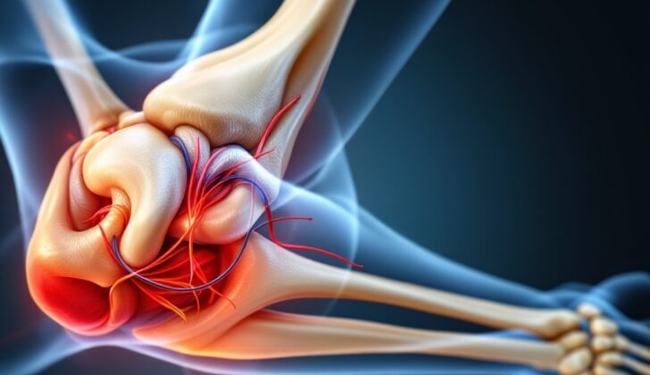
The medial epicondyle is a part of the bone where certain ligaments and muscles, specifically the ulnar collateral ligament and flexor-pronator mass, start. This group of muscles includes the pronator teres, flexor carpi radialis, palmaris longus, flexor digitorum superficialis, and flexor carpi ulnaris. These muscles have a blood supply from the superior and inferior ulnar collateral arteries. The ulnar collateral ligament, which starts at the medial epicondyle, plays an important role in keeping the elbow stable when stressed. The ulnar nerve, another crucial part of the elbow, runs behind and along the groove of the medial epicondyle. The medial epicondyle also happens to be the last bone in the elbow to mature, typically around ages 14 or 15. Therefore, it’s more likely to get injured in children.
When fractures happen to the medial epicondyle, they can vary in terms of how many pieces the bone breaks into and how much the bone moves from its usual place. Sometimes, when an elbow dislocates, the medial epicondyle can break as well, and pieces of it may get stuck in the elbow joint.
What Causes Medial Epicondylar Elbow Fractures?
A medial epicondyle fracture is often caused by physical stress on the elbow, like falling on an outstretched hand, or activities involving forceful throwing or wrestling. This type of fracture often goes hand in hand with dislocations of the elbow. Most of the time, the fracture results from the strain of certain muscles and ligaments attached to the medial epicondyle, particularly when the elbow experiences a sideways force, known as ‘valgus stress’. These muscles pull on the fractured part of the bone, causing it to move forwards. Sometimes, these fractures can also happen from a strong hit directly to the elbow.
Risk Factors and Frequency for Medial Epicondylar Elbow Fractures
Fractures of the medial epicondyle, a part of the elbow, make up 12% of all elbow fractures in children. These fractures are most often seen in children between 9 and 14 years old. Around 75% of these types of fractures happen in boys. The risk of these injuries increases due to more incidents of collisions, falls, and certain types of stress on the elbow, particularly in children who are active athletes.
- 12% of elbow fractures in kids are medial epicondyle fractures.
- These fractures are most common in children aged 9 to 14.
- Boys account for approximately 75% of these fractures.
- Increased instances of collisions, falls, and certain stresses on the elbow in active children could raise the risk of these injuries.
Signs and Symptoms of Medial Epicondylar Elbow Fractures
When someone suffers an acute injury, it is critical to gather detailed information about how the injury occurred and when it happened. The person’s medical history, including past injuries, and location of the current symptoms, also contribute to the evaluation. For athletes, it is important to know about their dominant hand, their sport, their training routines, and any recent changes in their activities. Understanding the patient’s daily requirements is vital in providing personalized care. Some patients may express concerns about restricted movement or neurological symptoms like decreased sensation or weakness. Any history of elbow instability, such as dislocation, should also be identified.
Physical examination plays an essential role in assessing the injury. The first step is to examine the skin for any cuts, swelling, bruising, deformities, or fluid accumulation. It also involves feeling the bony parts of the elbow for any tenderness and evaluating the range of movement. Patients often display sensitivity over the inner side of the elbow accompanied by swelling in the soft tissues. They may experience pain, a grating sensation with elbow movement, or a mechanical restriction to elbow motion. Additionally, checking the function of the ulnar nerve is an essential part of the examination. This assessment involves looking at the spreading and coming together of the fingers and preserved sensation in the little finger and the inner half of the ring finger. In addition, it is necessary to thoroughly examine the blood vessels and look for other fractures in the upper arm, including in the radius and ulna bones.
Testing for Medial Epicondylar Elbow Fractures
To check for medial epicondyle fractures, typically a type of elbow fracture, certain images of the injured area are needed. These images are taken from the front, the side, and at an angle (oblique) of the elbow using x-rays. The angled image is particularly helpful in determining whether parts of the bone have been moved out of their normal position.
If you look at the x-ray images, you can identify these fractures if the outline of the bone appears to be broken. Another clue can be a lack of smooth lines along certain parts of the bone. There is a particular piece of your bone, called the trochlea, that doesn’t usually show up on x-rays for children under eight. So if you can see it, this suggests that a piece of the fractured bone might be stuck in there.
Fractures like these can be categorized based on the Watson Jones classification which helps doctors decide on the best treatment plans. This system classifies the fractures into four types. Type I fractures are those, which have a displacement lesser than 5 mm without any rotation, and usually these are treated without surgery. On the other hand, Type II fractures have a displacement greater than 5mm and rotation in the bones.
Type III fractures have bone fragments trapped without the joint being displaced or moved from its usual location. In type IV fractures, the bone fragments are trapped and the joint is also dislocated. Usually, type III and IV fractures need surgical treatment. Type II fracture treatments are decided on a case-by-case basis considering the patient’s unique circumstances. In cases where the bone has gradually been damaged due to repetitive stress (stress reaction or stress fractures), typically in athletes who throw often, the usual treatment involves rest and avoiding activities that could worsen the injury.
Treatment Options for Medial Epicondylar Elbow Fractures
Non-Surgical Treatment
Best way to treat isolated medial epicondyle fractures that are non-dislocated is by avoiding surgery. This method involves a brief period of wearing a long arm cast during the first week, with the elbow bent to 90 degrees. After the splint is taken off, a protected workout program should start, which includes exercises to restore a full range of motion. Stress fractures are also generally managed without surgery.
Surgical Treatment
In some cases, surgery may be necessary to treat medial epicondyle fractures. This is especially true for open fractures, cases where pieces of the fracture get trapped within the joint, and when the elbow shows instability. Surgery is needed in about 5 to 18% of medial epicondylar fractures when fragments of the bone get stuck in the joint. The types of surgical techniques used include the use of open reduction and internal fixation with a screw and washer setup, Kirschner wires, sutures, and tension band techniques. K-wires are the most commonly used option for fragmented fractures in children. To get better access to the lower part of the upper arm bone, a back of the elbow approach is preferred. The patient might be placed either on their back or on their side with the hand placed on the back for better surgical access.
What else can Medial Epicondylar Elbow Fractures be?
- Fracture of the inner part of the elbow (known as a medial condylar fracture)
- Fracture above the elbow knob (known as a supracondylar fracture)
- Swelling and pain in the inner part of the elbow (medial epicondylitis or “golfers elbow”)
- Elbow coming out of its joint (elbow dislocation)
- Swelling at the back tip of the elbow (olecranon bursitis)
- Pressure on the nerve in the elbow causing pain (cubital tunnel syndrome)
- Injury to the ligament on the inside of your elbow (ulnar collateral ligament injury)
What to expect with Medial Epicondylar Elbow Fractures
Both surgical and non-surgical methods of treatment have been proven to result in good recovery outcomes. Research reviews show a success rate of 92.5% for healing bone (bony union) in cases of medial epicondylar fractures (a type of elbow fracture) that were managed with surgery. On the other hand, a lower success rate of 49.2% for healing bone was observed in cases managed without surgery.
With continued observation ranging from 2 to 5 years, studies suggest that non-surgical treatment is effective for medial epicondylar fractures that are minimally displaced (slightly out of normal position) and have a stable elbow condition.
Possible Complications When Diagnosed with Medial Epicondylar Elbow Fractures
Medial epicondyle fractures, which often occur in children, can lead to various complications. About 60% of these fractures are linked to dislocation of the elbow. Some children (around 10% to 15%) may also end up with ulnar nerve dysfunction due to such fractures. This means that they might face difficulty moving or feeling in their forearm and hand. Other possible complications from these fractures can be stiffness or instability in the elbow, deformity, delayed healing (nonunion), and wound infections. In some cases, serious infections of the joint (septic arthritis) or bone overgrowth in muscles (myositis ossificans) have been reported.
Based on extensive review studies, almost 10% of acute medial epicondyle fractures in children had ulnar nerve involvement. This can cause loss of sensation along the right side of the ring finger and across the small finger. Patients with this type of nerve issue (ulnar nerve palsy) typically need surgery, specifically ulnar nerve decompression. Research has shown that these patients can fully recover their nerve function after surgery, particularly if treated early.
Preventing Medial Epicondylar Elbow Fractures
Starting movement early after surgery to fix displaced medial epicondylar fractures, or fractures of a specific part of the elbow, has been shown to lead to improved results. After-surgery rehabilitation is really important for ongoing recovery. During this phase, the arm is kept in a fixed position, and patients start an exercise program. Doctors and physical therapists will guide patients through exercises to improve the movement range of their elbow and the rotation of their forearm. Once the fracture starts to heal, patients should start strength training exercises. This will help to speed up the recovery process and ensure it is as complete as possible.


