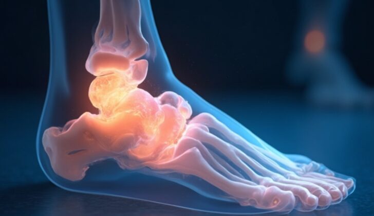
What is Talus Fracture?
The talus is the second largest bone in the foot, and it plays a critical role in foot and ankle movement because it connects the leg to the foot. The talus bone is rather unique and complex, with three distinct points where it connects to other bones. It’s made up of a head, neck, and body, and two-thirds of it is covered by a type of tissue called articular cartilage. In addition, it has few places where muscles or tendons attach.
The round part of the talus, known as the talar head, is covered by a type of cartilage called hyaline cartilage. It connects to the navicular bone at the front, linking the ankle and the middle part of the foot. It also connects to a bone called the calcaneus at the bottom. The body of the talus connects to the calcaneus from below and has two main points of connection. One of these points, the middle facet, is most commonly involved in a condition known as talocalcaneal coalition, accounting for 45% of cases. The upper part of the body of the talus connects to the tibia bone at the ankle. The neck of the talus, which connects the head and body, doesn’t have a surface for connecting with other joints or cartilage.
The back of the talus is made up of two bumps, or tubercles. A tendon known as the flexor hallucis longus tendon runs between these two tubercles. In some people, there’s a longer lateral tubercle known as a Stieda process, or a non-fused lateral tubercle ossification center known as os trigonum. Both of these are normal variants and can be involved in conditions ranging from fractures to os trigonum syndrome.
The side process of the talus bone connects to fibula at the top and forms a portion of the back subtalar joint. Fractures of this side process are often known as “snowboarder’s fractures,” and they’re frequently missed in initial x-ray scans. The best way to evaluate this process is through front-to-back (AP) x-ray scans of the ankle.
Blood supply to the talus comes from three arteries: the posterior tibial, dorsalis pedis, and perforating peroneal arteries. The blood supply is mainly outside the bone due to the extensive cartilage coverage, making it easily disrupted if fractures or dislocation occur, leading to bone death due to lack of blood supply.
What Causes Talus Fracture?
Head and neck fractures are often linked to high-energy accidents, like car crashes or serious falls. These types of incidents can also cause injuries to the body of the talus, which is a bone in the foot. However, sports injuries can also lead to bone injuries or breaks. For example, certain actions such as snowboarding or twisting the foot inwards or outwards can cause harm to the outer structure of the talus or result in the fracturing of its lateral process, which is a part in its side.
Risk Factors and Frequency for Talus Fracture
Talar fractures, or breaks in the talus bone in the foot, are rare. They make up less than 1% of all body fractures and only between 3% to 6% of foot fractures. These fractures can happen to people of all age groups, but are more common in younger people who are involved in car accidents. Also, these fractures are more common in men, with 73% of talar fractures happening to them. Their occurrence doesn’t seem to be increasing, but hospitals are seeing more of these injuries due to increased survival rates from serious accidents. On a related note, there’s a reported increase in a specific type of talar fractures – lateral process fractures – among snowboarders.
- Talar head fractures, the rarest type, account for 5 to 10% of talar fractures.
- Talar neck fractures, which were first noticed in World War 1 pilots, were once thought to be the most common, but recent studies suggest they make up only about 5% of talar fractures and are less common than body fractures.
- Understanding these fractures can sometimes be complicated because of differences in defining the locations of the neck and body of the talus bone.
- It’s worth noting that talar neck and body fractures often occur alongside fractures of the heel (calcaneal) and spine.
- The reported incidence of body fractures varies a lot, between 13% to 61% of talar fractures.
Signs and Symptoms of Talus Fracture
An injury to the ankle is typically followed by swelling, bruising, and sometimes, limited movement in the ankle and foot joints. This might make it difficult for individuals to put weight on the affected ankle. There are several types of fractures that can happen in the ankle region.
- Talar Head Fractures: These fractures cause pain, swelling, and tenderness at the top of the foot, and pain during movement.
- Lateral Process Fractures: Initially, these fractures may not be visible on X-rays, but if there is recurring pain on the side of the ankle after certain foot movements, they should be considered. If the lateral ankle pain doesn’t improve with standard treatments, evaluation for a lateral process fracture should be considered.
- Posterior Process Fractures: The “nutcracker sign” is associated with these injuries – pain and a grating sensation when the foot is bent downwards. Tenderness near the Achilles tendon and behind the talus bone in the ankle may also be signs.
- Pain with big toe movement: This may be a sign of an injury in the area as the flexor hallucis longus, a tendon that moves the big toe, runs adjacent to the talus bone.
However, fractures in other parts of the talus bone, a crucial component of the ankle, may not always have clear signs.
Testing for Talus Fracture
Diagnosing fractures in the foot doesn’t involve any laboratory tests. Instead, a series of x-ray angles are taken to provide clear images of the ankle and foot. These include anteroposterior and lateral views of the ankle, and in certain cases, some specialized views like the Canale or Harris radiographic views may be taken to view specific parts of the foot and ankle. These special views usually provides a clearer image of the fracture site, although they are often not used thanks to the wider availability of CT scans. For more detailed images and to aid with surgical planning, a multiplanar computed tomography (CT) scan can be carried out.
Fractures in the talus, the large bone in the ankle, are categorized based on whether they affect the head, neck, or body of the bone. Talar head fractures occur at the joint surface and may have related dislocation of the talus and fractures of surrounding bones. Talar neck fractures occur when there’s a break anterior or inferior to the lateral talar process and the talar dome cartilage. Depending on where these talar fractures are and their severity, they are further classified using the modified Hawkins-Canale system.
For instance, Type I fractures are those that are nondisplaced and are the least severe, so they pose the smallest risk of bone death (osteonecrosis). Type II fractures have displacement with related subluxation or dislocation of the subtalar joint. Type III fractures have displacement with subluxation or dislocation of the subtalar and tibiotalar joints, posing a high risk of osteonecrosis. Type IV is the most severe and involves dislocation or subluxation of the subtalar, tibiotalar, and talonavicular joints.
Talar body fractures involve breaks in the talar dome, lateral and posterior processes, and the body itself. These fractures are described using the Sneppen classification system based on the specific part of the talar body that is affected. For instance, fractures of the posterior process (Sneppen D) typically involve the lateral tubercle more often than the medial tubercle, while lateral process fractures (Sneppen E) frequently occur with forced dorsiflexion and external rotation or eversion of the foot.
Treatment Options for Talus Fracture
If a fracture in the talar head, a part of the ankle bone, doesn’t shift out of place (nondisplaced), it’s typically treated with non-surgical methods. However, if the fracture is out of place (displaced), surgery is required to realign the joint and prevent problems such as joint decay and bone death.
Fractures in the neck of the ankle bone fall into different categories. Type I fractures are usually treated without surgery. But if even a small shift is noticed in the position of the fracture, surgery may be needed for corrections. This highlights the importance of using CT scans to carefully evaluate these fractures. Type II fractures always need surgery for correction. Type III and IV fractures are initially managed by realigning the bone without surgery in the emergency room. This process helps to lessen the tension on the skin and reduce injury to the surrounding soft tissues. Surgery for a permanent correction is usually scheduled afterward.
Fractures in the body of the ankle bone may not need surgery if the fragments remain in their original position. But most of the time, these fractures are dislocated and require surgery to restore the correct alignment of the bone and joint. If the part of the ankle bone known as the posterior process is fractured, it is typically treated without surgery. However, if the discomfort continues even after conservative management, removing the fractured portion may become necessary.
Non-displaced fractures in the lateral process, another part of the ankle bone, are normally treated without surgery. However, if the bone fragments are too big or they have moved out of their original position, surgery will be required for correction. Extremely shattered fractures or fractures associated with joint injury may necessitate the removal of bone fragments. In general, surgeons opt for surgical correction for Type I fractures, removal for Type II fractures, and non-surgical management with no weight-bearing for Type III fractures.
What else can Talus Fracture be?
If someone has suffered a severe injury to their foot or ankle, there might be several possible injuries to consider. These could include broken bones in the foot and ankle, or damage to the soft tissues of the foot and ankle, such as the ligaments or tendons. Other structures that could potentially be injured include the nerves and blood vessels.
X-Ray images are often used to help determine the exact nature of the injury. Sometimes, people have extra bone fragments in their foot or ankle, which are a normal variation but can sometimes be confused with an injury. For example, the “os trigonum” is an extra bone that can look like a broken piece of bone from the back of the ankle.
However, an actual broken bone and an os trigonum can be distinguished by their appearance on an X-ray: a fresh break will look ragged and irregular, while an os trigonum will have smooth, clearly defined edges.
What to expect with Talus Fracture
Non-surgical treatment for talar head fractures that aren’t displaced, and proper surgical treatment for those that are displaced, usually result in excellent recovery results. The talar head, compared to the neck and body, has a better prognosis, largely due to its good blood supply.
When it comes to talar neck fractures, the type of fracture (classified by the Hawkins-Canale model) strongly impacts the risk of avascular necrosis, which is a condition where bone tissue dies due to lack of blood supply. Research notes that the risk of developing osteonecrosis and other conditions such as post-traumatic osteoarthritis increases with the degree of fracture displacement, the extent of injury to the surrounding soft tissue, and the success of surgical treatment. The rates of osteonecrosis in Types I, II, III, and IV fractures have been identified as 0 to 15%, 20 to 50%, 100%, and 100% respectively.
The ‘Hawkins sign’ is a specific feature on post-treatment images, showing a thin linear subcortical radiolucency, indicating that blood flow to the talar body is preserved. Stable and non-displaced defects of the talar dome often heal well, whereas displaced unstable fragments may suffer from osteonecrosis if not surgically treated.
Crush fractures of the talar body, where the bone is crushed or splintered, have the worst outcomes of all types of talar body fractures.
Possible Complications When Diagnosed with Talus Fracture
If a displaced fracture in the head or neck is not corrected, it could lead to secondary osteoarthrosis or necrosis, which are conditions that cause bone degradation or death. Interestingly, even if these types of fractures are treated correctly, there is nearly a 100% chance of developing osteonecrosis – bone death due to a lack of blood. This kind of bone death and secondary osteoarthrosis are common complications of broken talar bodies. If there are additional fractures of the head or neck, these can increase the chance for complications in body fractures. Crushed or pulverized talar body fractures have a high likelihood of bone loss and inadequate reduction, which leads to a higher risk of avascular necrosis, another form of bone death.
- Failure to correct displaced head or neck fractures can lead to secondary osteoarthrosis or necrosis
- Even with proper treatment, Type III and IV talar neck fractures may result in osteonecrosis
- Osteonecrosis and secondary osteoarthrosis are common complications of talar body fractures
- Additional fractures in the head or neck could increase the chances of complications in body fractures
- Crushed talar body fractures have a high likelihood of bone loss and problematic reduction
- Such fractures may result in higher risk of avascular necrosis


