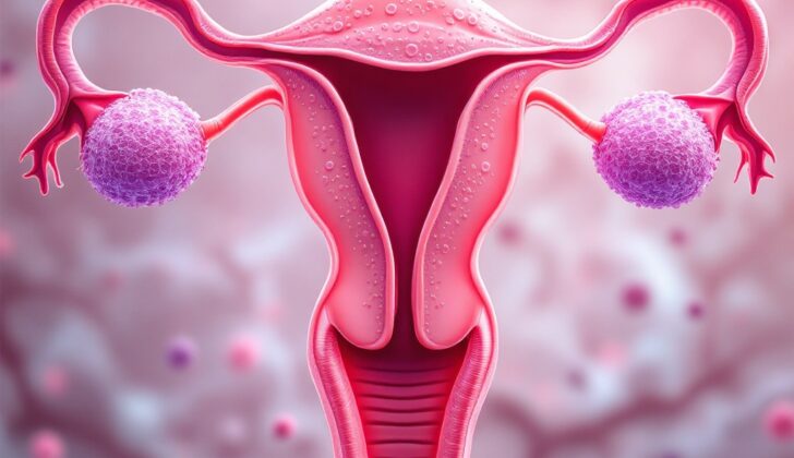
What is Premalignant Lesions of the Endometrium?
Endometrial cancer, or cancer that begins in the lining of the uterus, can be caused by certain abnormal medical conditions called premalignant endometrial lesions. One such condition is endometrial hyperplasia (EH), which is an overgrowth of cells in the endometrium. This can sometimes lead to endometrial cancer (EC).
Endometrial cancer is split into two types, each caused by a different precursor, or early-stage condition. The first type, known as endometrioid adenocarcinoma or Type 1 endometrial carcinoma, commonly develops from a condition known as Atypical Hyperplasia/Endometrioid Intraepithelial Neoplasia (AH/EIN). AH/EIN is an abnormal growth of cells in the uterus and is the early-stage condition for over 90% of endometrioid carcinomas. This type of cancer is usually diagnosed in women who are in their pre-menopause or peri-menopause phase. It’s mainly caused by an excess of the hormone estrogen in the body, combined with a lack of another hormone, progesterone.
Type 2 endometrial carcinoma, known as serous carcinoma, is often preceded by a condition called Endometrial Intraepithelial Carcinoma (EIC), which is another type of abnormal cell growth in the endometrium. This type of cancer is typically diagnosed in post-menopausal women and is associated with a thinning of the endometrium, known as atrophy.
Type 1 endometrial carcinoma makes up around 80-85% of all endometrial cancers. It shares many genetic mutations, or changes in the DNA, with invasive endometrioid carcinoma. Type 2, or serous carcinoma, makes up about 3-10% of all new diagnosed cases, and is driven by a specific genetic mutation known as P53 inactivation. There are also rarer types of endometrial cancer, like clear cell carcinoma, that account for the remaining cases.
What Causes Premalignant Lesions of the Endometrium?
The rates of endometrial cancer are rising globally, largely because of an aging population and increasing rates of obesity. Research agrees that there are changes within the cells (premalignant lesions) that happen years before invasive endometrial carcinoma develops. Atypical endometrial hyperplasia, an abnormal growth of cells in the lining of the uterus, can occur before endometrioid carcinoma, a type 1 endometrial cancer.
Here are some of the factors that can increase the likelihood of developing such abnormalities:
1. Being over 35 years old
2. Being of Caucasian race
3. Having a family history of endometrial hyperplasia or carcinoma
4. Experiencing early start of menstrual periods, lasting perimenopause, or late menopause
5. Being menopausal
6. Having related medical conditions like obesity, type 2 diabetes, and a certain type of ovarian tumor or a genetic predisposition to colon cancer
7. Exposure to certain treatments or hormones, like tamoxifen therapy, hormone replacement therapy (especially estrogen-only), or other hormonal exposures.
8. Environmental factors, like smoking, and certain genetic mutations can also increase this risk.
On the other hand, endometrial intraepithelial carcinoma (EIC), which precedes type 2 endometrial cancers, often progresses to more aggressive types. These types tend to be less dependent on estrogen and are often seen in postmenopausal women who have endometrial atrophy (thin uterus lining). This is in contrast to the hyperplasia seen in type 1 endometrial cancer.
Recent studies suggest that the loss of a tumor suppressor gene known as PTEN, although it is an important factor, is not enough to drive abnormal cell growth. The cells that lost PTEN also need to acquire additional molecular changes which contribute to formation of atypical hyperplasia. Since PTEN helps to regulate growth signals, its loss can cause over activity in these pathways, which can lead to overgrowth of certain cell types, such as those found in the uterine and breast tissue.
In fact, inactivation of PTEN is seen in over 70% of these type of lesions, making it an early contributor to atypical endometrial hyperplasia. Many other genetic changes have been identified through sequencing studies to drive type 1 endometrial carcinoma. This involves disruptions in a variety of different signalling pathways and genes. One gene worth noting is ARID1A, another tumor suppressor gene whose mutation can be observed.
For type 2 endometrial cancer, TP53 inactivation is a major contributing factor. Mutation or inactivation of TP53 is an early event in the development of these types of tumors and occurs in almost 75% of cases. Other genetic changes have been implicated, including mutation in other genes and overexpression of various other genes and proteins. Evidence shows disrupted PI3K pathways in these cases as well, mainly due to mutations in PIK3CA, along with mutations in PTEN and PIK3R1.
Risk Factors and Frequency for Premalignant Lesions of the Endometrium
Endometrial cancer, a common form of cancer that affects the lining of the womb, varies by race and ethnicity. The incidence is highest among non-Hispanic White women and lowest among Asian women. Black women, unfortunately, experience the worst survival rates. It’s important to note, though, that the same kind of data is not available for conditions that can lead to this cancer.
In countries like the US, endometrial cancer is the fourth most common cancer in women. The rates are continually increasing, especially in the age group of 40 to 44. According to predictions by the American Cancer Society, around 65,620 cases of uterine cancer will be diagnosed in 2020 in the US.
- Endometrial cancer is most common amongst non-Hispanic White women.
- Asian women have the lowest rates of endometrial cancer.
- The survival rates for endometrial cancer are worst among Black women.
- Endometrial cancer is now becoming more common amongst women aged 40 to 44.
- About 65,620 cases of this particular cancer are expected to be diagnosed in 2020 in the US.
Endometrial hyperplasia, a condition that leads to an increase in cells in the lining of the womb, tends to occur three times more often than endometrial cancer. If it’s not treated, it can develop into cancer. Research estimates suggest around 200,000 new cases of endometrial hyperplasia are diagnosed each year in developed countries, but this might be an underestimation.
One detailed study found that atypical endometrial hyperplasia, a more serious form of the condition, is 14 times more likely to turn into endometrial cancer compared to women with a certain abnormality in the womb lining but without hyperplasia. Women with endometrial hyperplasia without this more serious change in their cells are not likely to develop cancer.
- Endometrial hyperplasia is at least three times more common than endometrial cancer.
- Approximately 200,000 new cases of endometrial hyperplasia are reported every year in developed countries.
- Atypical endometrial hyperplasia can increase a woman’s risk of developing endometrial cancer.
- Endometrial hyperplasia without atypical cells is less likely to develop into cancer.
Signs and Symptoms of Premalignant Lesions of the Endometrium
Endometrial hyperplasia is a condition where the lining of the uterus becomes overly thick. Most of the time, people with this condition experiences unusual bleeding from the uterus. In women who can still have babies, this can manifest as heavy periods or bleeding between periods. For women who have been through menopause, it often shows up as bleeding after menopause. Sometimes, though, it can also cause a discharge from the vagina. Some people only find out they have it when a Pap smear shows abnormal cells from the glands or lining of the uterus.
To figure out whether the unusual bleeding is due to endometrial hyperplasia, a comprehensive health history and physical examination are necessary. It’s important to rule out other potential reasons for the bleeding or other symptoms. A detailed examination of the lower part of the female reproductive system, including the outer part of the genitals (vulva), vagina, cervix, uterus, and ovaries should be done. This exam should include a pelvic exam.
For people who are significantly overweight, pelvic exams can be difficult to do properly. In these cases, medical professionals may use ultrasound (a type of imaging that uses sound waves) of the pelvis to check for potential issues. This can help rule out problems with the ovaries and other possible causes of the symptoms, like growths in the uterus known as fibroids.
Testing for Premalignant Lesions of the Endometrium
If a doctor suspects that a patient may have endometrial hyperplasia, which is an overgrowth of the lining of the uterus, they may conduct an imaging test. This is typically done between the 5th and 10th day of the patient’s menstrual cycle. In diagnosing endometrial hyperplasia, the doctor may look at the thickness of the endometrium – the inner layer of the uterus. If it measures less than 6 mm in women before menopause, or less than 5 mm after menopause, it’s less likely that the person has endometrial hyperplasia. The endometrium could appear smooth or may have an irregular surface.
Another sign that could appear on the scan is cysts or fluid-filled sacs. However, ultrasound scans can’t reliably tell the difference between endometrial hyperplasia and endometrial carcinoma, a type of cancer that begins in the uterus. This is true unless the cancer has started to invade the muscle tissue of the uterus, also known as the myometrium.
If a woman is having abnormal bleeding, a pregnancy test should always be carried out to rule out pregnancy. Additionally, all patients with unusual vaginal bleeding, regardless of whether they are pre- or post-menopause, should have their blood checked for platelet count disorders or clotting problems. A complete blood count – a test that measures various components in the blood – should also be done, as it can indicate anemia which may result from excessive or chronic blood loss.
Doctors usually make a final diagnosis of endometrial hyperplasia via hysteroscopy and direct biopsy of the endometrium. Hysteroscopy is a procedure where a thin, telescope-like instrument is inserted into the uterus, and a biopsy involves taking and examining tissue samples. Ideally, these tests should be done when the patient isn’t taking progestin-based hormones, which are used to treat various conditions related to menstruation and fertility. If the patient is taking hormonal therapy, a tissue sample should be taken 2 to 4 weeks after they stop taking the medication.
In conclusion, diagnosing endometrial hyperplasia can involve a variety of tests, including ultrasounds, pregnancy tests, and blood tests to look for anemia and clotting disorders. The condition is often finally determined with a tissue sample taken from the inner lining of the uterus.
Treatment Options for Premalignant Lesions of the Endometrium
The American College of Obstetrics and Gynecologists (ACOG) recommends several steps for managing patients with premalignant endometrial lesions which are early signs of potential cancer in the lining of the womb. These include, checking for the presence of womb cancer, planning treatment to account for any delay in diagnosing hidden cancer, and preventing the progression to full womb cancer.
Treatments for premalignant endometrial lesions fall under two categories, surgical and non-surgical. The main surgical treatment is total hysterectomy, which involves removing the womb. This procedure is the preferred treatment. However, in younger women who wish to have children, a high dose of a hormone medication known as progestin may be used along with regular close monitoring.
Removal of the womb via total hysterectomy is currently regarded as the most effective treatment for premalignant lesions of the womb. ACOG recommends that, when appropriate, this operation provides a certain treatment and check for any existing hidden cancer. Other surgical procedures such as supracervical hysterectomy, morcellation, and endometrial ablation are not recommended because of concerns that hidden cancer might be missed.
Different forms of hysterectomy can occur, these may be through the abdomen, vagina, or with minimal invasion (such as using laparoscopic or robotic techniques). The chosen method depends on the extent of the planned surgery and the surgeon’s skills. Compared to an abdominal hysterectomy, a laparoscopic or vaginal hysterectomy usually results in less pain, earlier hospital discharge, and quicker recovery.
Non-surgical management might be offered to younger patients who wish to maintain their fertility, or patients who may be at increased risk of complications due to other health conditions. The goals of non-surgical treatment are complete clearance of disease, return to normal womb lining, and prevention of invasive cancer.
Non-surgical treatments typically involve hormone therapy. However, womb lining removal is not advised as the completeness of removal cannot be assessed and it may make subsequent follow-up examinations difficult. Hormone therapy mostly uses progestins or suppression of estrogen effects. A large proportion of premalignant lesions clear with high-dose progestin therapy.
Progesterone, also a hormone, counteracts the cell-growing effects of estrogens. It usually has mild side effects such as occasional swelling or digestive disturbances. Sometimes it may lead to blood clots. It can also be a reasonable choice for elderly patients with premalignant womb lining or low-grade cancer.
Regular and detailed checks of the womb lining are required to measure regression, maintenance, or progression of the lesions. For patients who receive non-surgical management in order to preserve fertility, they should follow up with hysteroscopy and direct womb lining sampling. No blood or tissue marker is available for follow up, and imaging modalities do not have the required specificity, so follow up with hysteroscopy and direct womb lining sampling is needed.
Approximately 30% of patients see no improvement from hormone therapy. This could be due to a decreased availability of progesterone receptors and changes in the programmed cell death signaling of the womb lining cells. Additionally, there might be a decrease in progesterone receptor numbers and activation of the transforming growth factor signaling pathway.
What else can Premalignant Lesions of the Endometrium be?
When diagnosing endometrial intraepithelial neoplasia (EIN), or atypical hyperplasia (AH), specialists need to consider other conditions that could potentially look similar. These conditions could lead to a misdiagnosis, being incorrectly labeled as conditions that involve excessive growth or crowding of cells. These crowded cells may be wrongly identified as potentially harmful or cancerous growths, known as premalignant or malignant lesions.
A study found that 77% of misdiagnoses turned out to be harmless (like growths in the womb lining), 19% were actually premalignant lesions, and 4% were endometrial cancer.
Some harmless conditions could also be mistaken for harmful or cancerous growths. These include:
- Mucinous metaplasia (a change in the type of cells in the womb lining)
- Papillary mucinous metaplasia (a certain kind of cell change in the womb lining)
- Endometrial polyps (growths in the womb lining)
If a person’s pregnancy history isn’t available, the Arias-Stella reaction (a response from cells due to hormonal changes during pregnancy) could be misdiagnosed as a potentially harmful growth. It can also be challenging to differentiate endometrial hyperplasia with secretory changes (a thickening of the womb lining) from an extremely packed and proliferative late secretory endometrium (a womb lining that is preparing to shed during menstruation).
What to expect with Premalignant Lesions of the Endometrium
Atypical hyperplasia (AH), a condition characterized by abnormal cell growth, has been found to develop into a specific type of cancer, endometrial carcinoma, in almost 29% of cases. The average time it takes for this development is usually around 4.1 years.
There’s also research indicating that AH may increase the risk of simultaneous invasive carcinoma, which is a cancer that has penetrated deeper into the tissue or spread to other organs. Recent studies have found that up to half of the women diagnosed with atypical cells have endometrial cancer detected when their uterus (hysterectomy specimens) is removed and examined.
On a brighter note, when endometrial cancer occurs along with hyperplasia, it’s usually less advanced and not as aggressive. This results in a higher 5-year survival rate, meaning that more patients are alive after five years of diagnosis compared to other types of endometrial cancer.
Possible Complications When Diagnosed with Premalignant Lesions of the Endometrium
Atypical hyperplasia can lead to severe complications such as intense bleeding from the uterus. This may be so severe that it requires immediate medical attention or surgery. In some cases, the bleeding is so intense that a blood transfusion may be necessary.
Common Complications:
- Severe bleeding from the uterus
- Need for immediate medical intervention or surgery
- Potential need for a blood transfusion
Preventing Premalignant Lesions of the Endometrium
Endometrial cancer (EC), a type of cancer that starts in the lining of the uterus, used to be mainly associated with women who had gone through menopause. However, with the rising issue of obesity in the U.S., we’re seeing more and more cases of endometrial hyperplasia (EH)/endometrial intraepithelial neoplasia (EIN), and cancer among younger women, including those who are still having periods or are in the stage before menopause.
This shift makes diagnosing and treating EH more challenging for doctors. EH is a condition where the lining of the uterus becomes too thick, which could lead to cancer. It’s important for doctors to intervene early on and educate patients about what they can do to lower their risk. This involves discussing changes in lifestyle that can help, like quitting smoking, improving diet, and becoming more active, all of which could help prevent the disease from developing.


