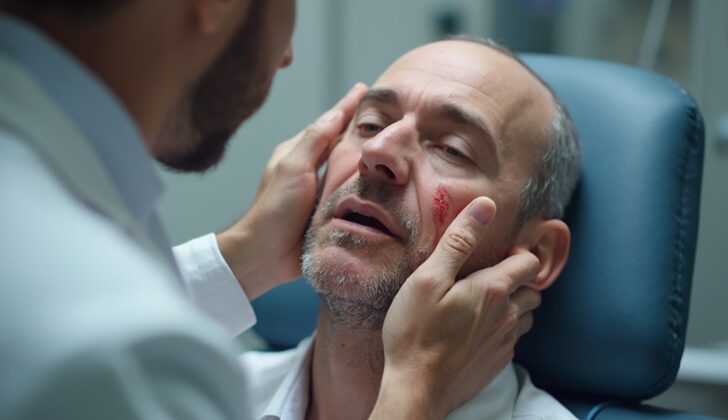
What is Maxillary Sinus Fracture?
Facial injuries are a common reason why people visit the emergency department. Injuries to the midface, or the central part of the face, are especially difficult for doctors to treat. Specialists like ENT (ear, nose, and throat) doctors and oral and jaw surgeons often assess and treat fractures in the maxillary sinus, an area within the midface. It’s important to understand how to diagnose and treat these fractures because they have a significant financial and emotional impact on the patient and the healthcare system. Patients with these injuries usually have fractures in multiple facial bones, which can require major surgery and a long recovery period.
Though the term ‘maxillary sinus fracture’ can refer to any fracture in this area, this explanation mainly focuses on fractures in the front and back walls of the maxillary sinus. Fractures in other parts of the midface are beyond the scope of this explanation.
To understand how to diagnose and treat maxillary sinus fractures better, we need to know about the structure of the midface. The midface is made up of several facial bones including the maxilla, zygoma, sphenoid, lacrimal, nasal, ethmoid, and palatine. A trauma that affects any of these bones might lead to a maxillary sinus fracture. This sinus, shaped like a pyramid, is the first one to form during development and is the largest sinus close to the nasal passage. It has various borders made up of different bones and also houses key nerves and blood vessels.
What Causes Maxillary Sinus Fracture?
Maxillary sinus fractures, or breaks in the bones around the upper areas of your cheeks, usually happen because of a strong blow to the face. The way people get these injuries can depend on things like their age, where and how the force hit them. This type of trauma often happens due to car accidents, domestic arguments, falls, workplace accidents, or attacks, sometimes involving a weapon.
Injuries to the middle part of the face, especially the side, are more common than injuries to the front part of the face. Interestingly, about 5% of lower jaw fractures can also cause isolated fractures around the back and side of the maxillary sinuses, the air-filled spaces within your cheeks.
In addition, it’s worth noting that almost half of the people with maxillary sinus fractures also have associated nerve injuries. The infraorbital nerve, which is responsible for feeling in the lower eyelid, upper lip, and cheek, is most commonly affected, followed by the facial nerve, which controls the muscles of facial expressions.
Risk Factors and Frequency for Maxillary Sinus Fracture
Understanding the prevalence of Maxillary Sinus Fractures (MSFs) is challenging, because many different facial bones can be involved. A lot of patients with MSFs have several fractures in different facial bones, all of which might impact the maxillary sinus. People with facial bone fractures are mainly males between the ages of 21 to 30. The most commonly fractured facial bone is the nasal bone, but patients with this type of fracture usually don’t need surgery. However, fractures to the mandible, or jawbone, which makes up a part of the facial structure, often require surgery and account for 38 to 63% of all cases.
- The prevalence of MSFs is difficult to estimate due to the complexity of the facial bone structure.
- Patients with MSFs often have multiple fractures that can affect the maxillary sinus.
- Facial bone fractures are most common in males aged 21 to 30.
- The nasal bone is frequently fractured, but usually doesn’t require surgery.
- The most common bone requiring surgery when fractured is the mandible, representing 38 to 63% of surgery-required fractures.
- Mandible fractures can be associated with posterior maxillary sinus fractures in up to 11% of cases.
- The second and third most common bones to be fractured are the maxilla and the orbit, both part of the maxillary sinus structure.
Signs and Symptoms of Maxillary Sinus Fracture
When dealing with patients who have sustained potentially traumatic injuries, it’s crucial to gather as much information as possible. This could include the patient’s age, details about how the injury happened, and how long ago the injury took place. Since the patient might be unable to communicate due to the injury or being on medical support measures like a breathing tube, doctors often rely on information from bystanders, family, emergency room staff, and emergency responders.
Furthermore, conducting a physical examination in such scenarios can be challenging due to factors like significant facial swelling or the presence of drugs and alcohol.
The evaluation process follows the Advanced Trauma Life Support protocol, which focuses on assessing the patient’s ability to breathe and circulate blood, as well as looking for any neurological issues. A Glasgow Coma Scale Score is calculated for this purpose, with an importance also given to examining for nerve function issues or suspected head injuries.
Once the patient is stable and a basic history has been taken, a comprehensive head and neck examination is conducted. Here is a breakdown of the examination process:
- The entire head and neck should be checked for any visible wounds, bruises, or active bleeding.
- Observe for any deformities, especially in the nose, cheekbone area, and jawbone.
- Try to control any bleeding from cuts, clean them out, and close them if possible.
- Feel the facial bones to evaluate for any irregularities or instability.
- Perform a detailed oral examination as there may be bones poking through into the mouth.
- Look for any loose teeth or bite changes, which might suggest a jaw fracture.
- Even though a nasal examination might be limited, check for fluid leaks, blood clots, or pus.
- Examine the ears for signs such as Battle’s sign, blood collections, fluid leakage, or blood behind the eardrum suggesting further damage.
Special attention is given to a neurological examination since nerve damage could indicate the need for surgery. Tests include measuring pupil size and response to light, extraocular muscle function tests, vision tests, and checks for signs of facial nerve damage. The doctor should also be aware of possible “raccoon eyes” suggesting further head trauma and corneal abrasions visible on slit-lamp examination.
Testing for Maxillary Sinus Fracture
If a doctor suspects maxillofacial fractures (facial bone injuries), they might rely on physical examination first. However, often they would need more detailed information that can be provided by various imaging techniques. These imaging techniques can help recognize any additional injuries, aid in pre-surgery planning, and give an idea about future treatment steps.
Among different imaging methods, computed tomography, also known as CT scan, without contrast is widely used in facial trauma. This is because a CT scan can quickly show broken bones and the extent of displacement, which is vital information for deciding further treatment options. CT scans can also identify related fractures, foreign bodies, local bleeding, and some soft tissue injuries. Plus, by offering a three-dimensional perspective (3D-CT), CT scans help doctors better understand the complexity of fractures. However, because two-dimensional CT scans offer excellent bone visuals and have lower costs and radiation levels, 3D-CT scans are usually recommended only for severe multiple facial injuries and before and after surgery.
Other less-commonly used imaging techniques include magnetic resonance imaging (MRI), plain x-rays, and ultrasound. While these techniques have limited use compared to non-contrast CT scans, MRI can provide supplementary information especially in case of suspected cerebrospinal fluid leaks and eye tissue injuries. Furthermore, it can differentiate between herniated orbital fat and entrapped muscle. Ultrasound can be used to diagnose superficial fractures specifically in the zygoma (cheekbone) and nasal bones, but its usefulness is limited as it is unable to accurately diagnose non-displaced fractures or show the floor of the eye socket due to its poor penetration capability.
Treatment Options for Maxillary Sinus Fracture
In cases of facial trauma that result in maxillary sinus fractures (MSFs), the first priority is to stabilize the patient. This includes ensuring they have a clear airway, controlling any bleeding, and maintaining their heart functions. Sometimes, a preemptive tracheostomy (a procedure to create an airway) is necessary. With most facial fractures, immediate surgery isn’t usually needed and treatment can be given up to two weeks after the injury. However, most patients will probably need to stay in the hospital.
The approach to treating maxillary sinus fractures isn’t clear cut and experts differ on whether to opt for surgery or a non-surgical route. Those in favor of non-surgical treatments often highlight the potential risks and complications of surgery, arguing that conservative treatment might be a safer option. These conservative treatments include antibiotic use to prevent sinus infection, steroids to reduce swelling, and regular follow-ups. The use of antibiotics in these situations, however, is a point of debate.
One study compared patients with non-operated maxillary sinus fractures and concluded that a short course of antibiotics did not effectively prevent infection. In 2016, another study found no difference in infection rates between patients who did not receive antibiotics and those who did, regardless of short or long-term use.
When it comes to surgery, the factors deciding whether it’s necessary for MSFs are much like those for LeFort fractures. These factors include noticeable facial distortions, the need to restore the facial structure, correcting irregularities in the bite, and repairing the forward protruding part of the face.
If surgery is chosen, the typical method involves repositioning the fractured segments and securing them with plates, screws, mesh grafts, or dissolvable foils. How the surgeon decides to carry out this procedure can vary.
In recent years, new techniques have been introduced, although none has been proven superior. In 2017, three different groups reported using urinary balloon catheters and/or natural protein glue to hold the fractured segments in place. Yang and a group of colleagues developed a new process to reduce fractures using an endoscope (a tool used to examine the interior of a body organ) and ultrasound-guided nasal reduction of the maxillary sinus wall.
What else can Maxillary Sinus Fracture be?
The broken bones in the face can include:
- Zygomatic arch (cheekbone) fracture
- Mandible (jawbone) fracture
- Fracture in the lower edge of the eye socket (inferior orbital rim)
- Floor of the eye socket fracture (orbital floor)
- Nasal bone fracture (broken nose)
- Nasoorbitoethmoid fracture, a complex fracture that involves the nose, the corner of the eye, and the thin bone at the front of the skull
Other conditions related to the nose and facial area can also include:
- Acute, chronic, or recurrent sinusitis in the maxillary sinus (rhinosinusitis, an inflammation or infection of the sinuses)
- Facial hematoma or bruise (blood collecting under the skin following an injury)
What to expect with Maxillary Sinus Fracture
The outlook for patients with maxillary sinus fractures often depends on how severe the injuries are. Since those with such fractures often have other injuries as well, predicting their recovery can be tricky. Certain factors like loss of smell, complete vision loss, severe shift of the fracture, and delayed treatment signal poor prognosis. However, these symptoms usually indicate a more serious injury than just a maxillary sinus fracture.
The most frequent long-term complications include dental issues with alignment or incorrect healing of the fracture site.
Possible Complications When Diagnosed with Maxillary Sinus Fracture
Several complications have been mentioned in medical literature, such as:
- Entrapment of extraocular eye muscles (specifically the inferior rectus and inferior oblique muscles)
- Infection of the eye socket (orbital cellulitis)
- Abscess (pus-filled swelling) in the eye socket
- Air trapped in the eye socket (orbital emphysema)
- Sunken eye (enophthalmos)
- Lower than normal eye position (hypophthalmos)
- Maxillary sinusitis (inflammation of the sinuses)
- Pouch-like formation filled with mucus in the sinus (maxillary sinus mucocele)
- Nosebleed (epistaxis)
- Leakage of cerebrospinal fluid from nose (CSF rhinorrhea)
- Fistula (an abnormal connection) between the mouth and maxillary sinus
- Improper fitting of the teeth (dental malocclusion)
- Uneven appearance of the face (facial asymmetry)
- Aesthetic concerns
- Sensitivity to pressure
- Chronic facial pain
- Numbness in the lower eye area (infraorbital nerve paresthesia)
Preventing Maxillary Sinus Fracture
Maxillary sinus fractures, or breaks in the upper jaw bone, are typically caused by unexpected trauma, making them hard to prevent. However, taking several safety measures can help lessen the severity of these injuries. Some of these precautions include safer practices with vehicles and machinery.
Several experts suggest that actions such as installing traffic cameras, bettering road designs, adding speed bumps, and creating safer pedestrian routes can decrease the number of car accidents. This could help reduce these types of injuries since car accidents are a common cause.
In addition, using suitable protective gear during sports can lower the chances of getting injured on the field. Furthermore, steering clear of dangerous situations or locations can also help prevent injuries from assaults or falls.


