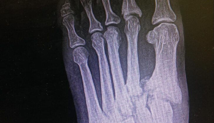
What is Lisfranc Dislocation?
The Lisfranc joint is the area where the metatarsal bones (bones in your feet labeled M1 to M5), and the tarsal bones (bones in your ankles labeled C1 to C3 and the cube-shaped bone) meet. The Lisfranc ligament, made up of three components, connects the innermost ankle bone (C1) to the second bone in your foot (M2).
You can damage the Lisfranc joint through a sprain, slight shifting of the bones, widening of the joint with or without breaking a bone, dislocation, or by a severe crushing injury. Lisfranc injuries are rare, affecting one in 55,000 people in the United States. These injuries are often wrongly diagnosed or improperly handled. Initial diagnosis misses around 20% of these injuries. Incorrect management of Lisfranc injuries can lead to severe complications like foot arthritis, chronic pain, and instability while walking.
What Causes Lisfranc Dislocation?
Lisfranc injuries can come from high-energy impacts like car accidents or falls from great heights, as well as low-energy events such as sports incidents. Depending on the intensity, these injuries are categorized differently.
High-energy Lisfranc injuries result from intense impacts like car crashes or falls, where the force crushes the foot, leading to serious immediate complications like open fractures, compartment syndrome, wounds opening up, or blood vessel disruption. These injuries don’t have typical signs that make them easy to identify.
On the other hand, low-energy Lisfranc injuries commonly occur during sports, when the foot is pointed and experiences a twisting or axial force. Pointing the foot weakens the ligaments at the top of the foot, making the base of the toes and the capsule on the bottom of the foot susceptible to twisting injuries. This type of injury is common in sports like basketball, football, and rugby and can even happen to horseback riders, which could result in a sideways dislocation of the toes.
Research has shown that certain physical characteristics may make people more prone to Lisfranc injuries and the complications that follow. For instance, one study found that more than half of the patients with Lisfranc injuries had a smaller ratio between the length of the second toe and the length of the foot. Another study revealed that a shorter height of the joint between the second toe and the foot increased the risk of a serious Lisfranc injury.
Risk Factors and Frequency for Lisfranc Dislocation
Lisfranc injuries are not very common. They make up only 0.2% of all fractures. However, their actual incidence could be higher because about 20% go undiagnosed. It is estimated that about 1 person in every 55,000 gets a Lisfranc injury each year. Also, for every 60,000 to 88,000 people in a region, there is usually one case of Lisfranc injury reported in a hospital.
This kind of injury can happen to people of any age, but they occur mostly in people’s third decade of life. They are 2 to 4 times more common in males, although females are more likely to develop more serious, or unstable, injuries. Athletes are also more likely to experience these injuries, and there has been an increase in reports from this group thanks to advanced athletic training.
- Most Lisfranc injuries are caused by high-energy incidents like:
- Motor vehicle crashes, which account for 43% of cases.
- Falls from heights, making up 24% of cases.
- Crush injuries, contributing to 13%.
- Sports injuries, causing around 10% of the cases.
Signs and Symptoms of Lisfranc Dislocation
Patients usually come to the doctor with pain in the middle part of their foot, especially after an injury. The pain gets worse when they put weight on it or flex their foot to take a step or start to run. The injury can happen from an intense or minor event, but people often underestimate how serious it is at first. Some people don’t get medical attention right away and only see a doctor when complications start to show. If the pain in the middle of the foot continues for more than 5 days, is accompanied by swelling, and persistently changes the way the patient walks, a Lisfranc injury is highly suspected.
When the doctor examines the foot, they may notice swelling and tenderness in the middle part of the foot, as well as possible bruising on the sole of the foot. The swelling and soreness are often more noticeable over the joints where the bones in the middle part of the foot connect to the longer foot bones. In some cases, the foot may appear wider and flatter than before. The doctor may also notice a larger gap than normal between the first and second toes, a sign of instability in the intercuneiform (foot) bones.
Further examinations may be carried out by the doctor. For instance, if the patient straightens the foot and moves it side to side under weight, the pain might be reproduced. By individually checking the stability of the joints where the middle foot bones join the longer foot bones, or using what is known as the piano key test, the doctor is able to reproduce the pain and identify instability. A single-leg toe rise test is another way to point out instability in the Lisfranc joint when the patient is putting weight on the foot. It is also important for the doctor to ensure that the injury is not open, as open fractures or dislocations require immediate surgical treatment.
Testing for Lisfranc Dislocation
If you have a suspected Lisfranc injury, which affects the middle part of your foot, your doctor may take x-rays of both your feet while you are standing up. This includes a front view x-ray, a side view x-ray, and a specially angled x-ray. Standing x-rays are important because they can often reveal Lisfranc injuries that other x-rays might miss. In fact, up to half the time, key signs of a Lisfranc injury can be overlooked if you’re not putting any weight on your feet during the x-ray. It’s also important to take an x-ray of your uninjured foot at the same time so the doctor can compare the two and spot any differences.
A few things your doctor may look for in the x-rays to confirm a Lisfranc injury include signs of misalignment between certain bones, the presence of a tiny bone fragment (known as a fleck sign) which may suggest a torn Lisfranc ligament, spacing of more than 2 mm between certain bones, or a vertical displacement of the foot’s bones.
Sometimes, though, a standard x-ray isn’t enough to identify a Lisfranc injury. It can miss about 20% of these injuries. So your doctor might order a CT scan to get a better, more detailed look at your foot and identify more subtle signs of damage. A CT scan would be particularly useful in identifying smaller fractures or tiny displacements not clearly visible on a standard x-ray. It can also help guide the treatment plan.
Magnetic Resonance Imaging or MRI can be used to assess the extent of injury to the soft tissues and ligaments around the affected area. For an MRI scan, image sequences should be ordered in sagittal (front-to-back view), long axial (side view), and oblique coronal (tilted front view) planes.
Treatment Options for Lisfranc Dislocation
Lisfranc injuries, which affect the feet, can be treated either with surgery or non-surgery. These two options depend on how severe the injury is. The main goal of any treatment is to make sure the foot stability is restored, that long-term complications are minimized, and that the foot returns to normal function and movement.
Non-Surgical Treatment for Lisfranc Injuries
In some cases, doctors recommend a non-surgical treatment. This usually applies to injuries that haven’t caused any displacement or aren’t too unstable and can be seen under weight-bearing x-rays. It also applies to foot sprains without any bone injury or to patients who are not fit for surgery.
This non-surgical treatment involves reducing the injury, applying a splint, and raising the foot. The injured foot is placed in a short-leg cast or boot. After two weeks, the patient is checked again and x-rayed to make sure there is no displacement requiring surgery. During this period, the patient should avoid putting any weight on the injured foot.
After six weeks, if the symptoms have gone and the x-rays show improvements, it means that the patient can start putting weight on the foot again and start physical therapy. The patient can gradually start doing more physical activity using support to help carry the weight on the inside of the foot.
If after six weeks the patient is still feeling some pain but the x-rays show no displacement, more time in a boot or cast is required, up to four extra weeks, before starting physical therapy. In cases where the ligaments are disrupted, the foot might have to stay immobile for up to four months.
Most patients recover in two to four months, but some may take up to six months.
Surgical Treatment for Lisfranc Injuries
Surgery is the chosen treatment when there’s instability in the Lisfranc joint, when there’s a compartment syndrome, or an open fracture. A compartment syndrome is considered a medical emergency and must be checked immediately so a surgical cut can be done to alleviate pressure. Open fractures also need immediate surgery because they can cause serious infections. If these conditions are not present, the foot can first be closed, reduced, and immobilized until the swelling goes down and the surgery can be performed.
The surgery involves two parts. First, the foot is reduced (its normal alignment is restored) in a closed way or with minimal invasion, keeping it immobilized with a pin or external fixation. Then, it is permanently fixed through open reduction (putting the displaced bones back into their normal position), arthrodesis (the fusion of bones to relieve pain and restore stability), or tendon grafting. If a two-stage surgery is not necessary, the surgery is delayed for 10 to 14 days until the swelling is gone.
There’s always been a lot of discussion about the surgical procedures to treat Lisfranc injuries. There are several methods:
ORIF (open reduction internal fixation), which might involve the following:
* Placing a screw through the joint. This is a traditional low-cost solution that avoids damaging soft tissue too much. However, a single 3.5-mm screw through the joint might end up damaging it.
* Dorsal-bridge plating. This technique results in better functional outcomes than placing a screw through the joint. It’s useful for treating injuries with a lot of damaged fragments.
* Suture button. This newer technique has shown promising results but more research is needed to confirm its effectiveness.
PA (primary arthrodesis). Many patients who underwent ORIF developed post-traumatic osteoarthritis, which can then be treated with PA. Compared to ORIF with a screw through the joint, PA resulted in better function scores, activity levels after surgery, and a lower rate of complications. However, a recent review showed no significant difference in returning to work or activity, complications, or satisfaction rates. Although there’s confusion in the research, PA is usually the method of choice for Lisfranc injuries more than six weeks old, which are more likely to develop post-traumatic osteoarthritis.
Tendon graft reconstructive surgery is used for injuries where only the ligaments are affected. The evidence supporting this technique is quite limited and comes from studies on cadavers or small case reports.
What else can Lisfranc Dislocation be?
The most common mix-up when diagnosing a Lisfranc injury is with a simple midfoot sprain which is a less severe condition. This mix-up often leads to improper treatment. Some other conditions that may be mistaken for a Lisfranc injury due to similar symptoms include flat feet (known as primary pes planus), issues with the tendon in the lower leg (posterior tibial tendon dysfunction), and joint wear and tear (osteoarthritis). Each of these conditions requires different treatments, which makes correct diagnosis crucial.
What to expect with Lisfranc Dislocation
Injuries caused by high energy impacts tend to have a bad outlook and worse results. Also, if surgery is delayed for more than six months, the functional results tend not to be as good. Type B injuries, in particular, usually have a worse outlook than Types A and C because they are often discovered too late.
When it comes to surgical techniques for fixing the Lisfranc joint, the PA method tends to have the best outlook. This is because it doesn’t require another procedure or the removal of an implant later on. Plus, patients typically get back to their normal activities faster after this procedure compared to an ORIF.
Possible Complications When Diagnosed with Lisfranc Dislocation
After a Lisfranc injury, there can be both immediate and long-term complications. Acute, or immediate, complications may include something called compartment syndrome, infection, blockage of blood vessels (thrombosis), and wounds. Long-term, or chronic, complications may include issues with surgical hardware, flat foot (pes planus), and most frequently, posttraumatic osteoarthritis, a type of arthritis that develops after an injury. As many as 60% of patients may experience this form of arthritis.
Common Complications:
- Compartment syndrome
- Infection
- Thrombosis or blood vessel blockage
- Wounds
- Issues with surgical hardware
- Flat foot or pes planus
- Posttraumatic osteoarthritis
Recovery from Lisfranc Dislocation
Patients should avoid exercises that require weight-bearing for a period of 6 to 8 weeks post-surgery. Once this period is over, doctors will check if these patients are fit to walk using a boot or short-leg cast, which they have to use for an additional 6 weeks. By the time the third month comes around, most patients who have undergone Lisfranc joint surgery usually show good recovery and are fit enough to wear supportive shoes and begin physical therapy.
Preventing Lisfranc Dislocation
People who take part in activities that make them more prone to Lisfranc joint injuries should be aware of the following recommendations:
* Wearing the right shoes: Injuries to the Lisfranc joint that occur during sports or in specific jobs can be avoided by wearing specially designed footwear. Such shoes provide support and protection to the middle of the foot, which can be beneficial for athletes during their activities.
* Increasing training intensity slowly: By gradually intensifying their training routines, individuals can properly condition, strengthen, and improve the flexibility of their feet and ankles. This helps decrease the chance of injuries.
* Practicing proper movement techniques: Learning and applying the right techniques can help individuals reduce falls and awkward foot positions.
* Thorough warm-ups and stretchings before physical activity: These actions ready the muscles, tendons, and ligaments for movement and help to minimize the risk of injuries.
* Securing environmental safety: This involves pointing out any environmental hazards which might increase the likelihood of Lisfranc injuries. Following work guidelines carefully can also prevent accidents.
* Keeping strong and flexible: Exercises that maintain the strength, stability, and flexibility of the foot and ankle improve movement dynamics and help avoid injuries.
Coaches, trainers, and healthcare providers should endorse these preventative measures. They should also keep reminding athletes and workers need to seek help immediately if they get hurt.


