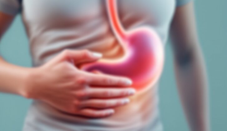
What is Gallbladder Mucocele (Hydrops of the Gallbladder)?
A mucocele of the gallbladder happens when there’s a long-term blockage in the tube that helps in bile flow, usually due to a stuck gallstone. This condition is also known as hydrops of the gallbladder. Most of the time, it’s not discovered until surgery, where it’s incidentally found during either a keyhole surgery or open gallbladder removal surgery. The detection of this condition is confirmed during surgery when, instead of the typical green or brown bile, the gallbladder is found to be filled with a clear mucus-like fluid. Usually, the patients show symptoms of acute or ongoing inflammation of the gallbladder.
What Causes Gallbladder Mucocele (Hydrops of the Gallbladder)?
Cholecystitis, or inflammation of the gallbladder, is caused by a malfunctioning gallbladder. The liver produces a fluid called bile, which travels down a tube called the bile duct and gets stored in the gallbladder. After you eat certain kinds of foods, particularly spicy or greasy foods, the gallbladder releases the stored bile. It travels through another tube called the cystic duct, back into the bile duct, and finally into a part of the stomach called the duodenum. This process helps with digestion.
If the gallbladder doesn’t work properly, it might not empty all of the bile, which can lead to the formation of gallstones. Gallstones can block the cystic duct. Additionally, the gallbladder also plays a role in concentrating bile and absorbing water from it. If any blockage prevents the gallbladder from emptying correctly, it can expand, storing up to about 1.5 litres of bile.
If this blockage is complete, usually due to a large gallstone in the cystic duct, the bile gets slowly absorbed by the lining of the gallbladder and gets replaced by a clear, watery mucus. This creates a condition called a gallbladder mucocele. Gallbladder mucoceles can also be caused by things that block the cystic duct or the lower part of the bile duct tree. This includes growths like polyps or tumors. Other conditions, like congenital strictures (birth defects that cause narrowing), gallbladder parasites, or pressure from liver disease or tumors, can also cause gallbladder mucoceles.
There are other factors that could lead to this problem. These include anything that increases the chance of getting cholecystitis, like drastic weight loss, prolonged use of total parenteral nutrition (TPN), stomach surgeries that disrupt the vagus nerve (which controls digestive tract function), severe illness, diabetes, high cholesterol, high calcium levels, and liver conditions like Caroli disease.
People with a condition called acute acalculous cholecystitis, inflammation of the gallbladder without any identifiable gallstones, can also form gallbladder mucoceles. Even though gallstones aren’t present, this still results in a stretched-out gallbladder because bile can’t be adequately released and is ultimately exchanged for a biliary mucus.
Risk Factors and Frequency for Gallbladder Mucocele (Hydrops of the Gallbladder)
Mucoceles, which can appear in individuals suffering from acute calculous or acalculous cholecystitis, are linked to gallbladder disease. This health problem can affect both men and women, but certain groups are more likely to experience it. Specifically, the risk of gallbladder disease rises for women, those who are overweight, pregnant women, and people in their 40s. Quick weight loss or sudden illnesses might also increase the risk, as can genetic predisposition and the formation of gallstones. Diseases like Sickle Cell disease that result in the breakdown of blood cells can further raise the chances of gallstone development. Despite their frequency, the majority of gallstones don’t present symptoms.
- In the United States, about 14 million men and 6 million women aged between 20 to 74 have gallstones.
- The chance of having gallstones gets higher as a person gets older.
- Being obese can elevate the risk of gallstones, especially in women because it increases the secretion of cholesterol in the bile.
- Those who have lost weight dramatically or are fasting are also at risk due to a condition known as biliary stasis which causes the bile in the gallbladder to become stagnant.
- Hormones play a role too; estrogen boosts cholesterol in the bile and decreases gallbladder movement. As a result, women of child-bearing age or those taking birth control with estrogen have a doubled risk of forming gallstones compared to men.
- People suffering from chronic diseases like diabetes experience increased gallstone formation and reduced gallbladder function due to nerve damage.
Signs and Symptoms of Gallbladder Mucocele (Hydrops of the Gallbladder)
Chronic cholecystitis is a condition that’s often characterized by progressive pain in the upper right part of the stomach, sometimes paired with bloating, intolerance to certain foods (especially fatty or spicy options), increase in gas, and experiencing nausea and vomiting. This pain could also extend to the middle of the back or the shoulder, and it could persist for years before a diagnosis is established. Acute cholecystitis, on the other hand, has similar symptoms, but they tend to be more severe. They can sometimes be mistaken for heart-related problems.
The presence of pain in the upper right part of the stomach when probed deeply, otherwise known as the Murphy sign, is a common indicator of this disease. Particularly in thinner people, the gallbladder can usually be felt at the right edge of the ribcage. In cases of extreme swelling, it can sometimes even be felt as low as the belly button. Patients often share history of a specific meal, like eating greasy foods such as pork chops and gravy, triggering an acute attack. These patients typically exhibit symptoms that have been gradually worsening over several days.
- Progressive upper right abdomen pain
- Bloating
- Intolerance to certain foods (especially fatty or spicy ones)
- Increase in gas
- Nausea and vomiting
- Pain that might extend to the middle of the back or the shoulder
- Presence of severe symptoms (in case of acute cholecystitis)
- Symptoms mistaken for heart-related problems
- Presence of the Murphy sign when the stomach is probed deeply
- Ability to feel the gallbladder at the right edge of the ribcage or as low as the belly button (in cases of extreme swelling)
- History of a specific meal triggering an acute attack
- Gradually worsening symptoms over a period of several days.
Testing for Gallbladder Mucocele (Hydrops of the Gallbladder)
To diagnose a gallbladder condition called cholecystitis, your doctor will likely perform a physical exam and take a detailed history of your health. Additionally, they’ll want to run some tests on your blood. They’ll look at the different components of your blood, like your white blood cells and other chemicals, through tests called a complete blood count and a comprehensive metabolic panel. The levels of these components might be normal or they might be high, especially in acute or severe cases of cholecystitis.
If your white blood cell count is high, it could mean there’s inflammation going on in your body. Similarly, higher numbers on the liver portion of your blood tests may point to an issue with your gallbladder. More specifically, a certain chemical called bilirubin, a waste product processed by your liver, might be high, which could mean there’s a blockage in the pipes that help your liver and gallbladder work properly, due to a stone, for example.
In severe gallbladder disease, these lab results might still fall within the normal range. If the liver, pancreas, and gallbladder are involved, your healthcare provider may want to further investigate with two more tests, checking amylase and lipase levels, to exclude inflammation of the pancreas, known as pancreatitis.
Medical imaging, like CT scans or ultrasounds, might also be used to get a better idea of what’s happening in your body. CT scans are often the first step here. These allow the doctors to see inside your body, and they might find signs of cholecystitis or gallstones this way. Ultrasounds are the best imaging test to use when looking at the gallbladder initially. With this, they might see a visibly swollen and angry-looking gallbladder with gallstones, or a stone that’s stuck and not moving in certain parts of the gallbladder.
In cases of acute cholecystitis, a specific scan called a HIDA scan is recommended. This is a special kind of scan that looks at how your gallbladder is functioning, and whether its outlet pipe is blocked. If there are no gallstones, but your symptoms suggest gallbladder problems, the doctors might use a chemical called cholecystokinin to help them diagnose a tricky kind of cholecystitis that doesn’t involve gallstones. People with this condition often have a low gallbladder “ejection fraction”, a measure of how well the gallbladder is functioning, with very severe cases showing 0%.
Treatment Options for Gallbladder Mucocele (Hydrops of the Gallbladder)
The suggested treatment for cholecystitis with hydrops, a condition where the gallbladder is enlarged due to fluid accumulation, is a surgical procedure called laparoscopic cholecystectomy. It’s favored because it typically involves fewer health risks and faster healing times. Essentially, this is a minimal invasive surgery where the doctor removes the gallbladder through small incisions in the abdomen using a tool called a laparoscope.
However, if for some reason, the patient is not suitable for laparoscopic surgery, the gallbladder can also be removed through an open surgery, which involves a larger incision. This method is used only if the patient’s condition or overall health makes the laparoscopic method unsafe or impractical.
If the patient is quite unwell and determined not to be a good candidate for surgery, they may be treated with a different method. This involves a procedure called percutaneous drainage, which means the doctors insert a small, flexible tube through the skin and into the gallbladder to drain the built-up fluid. This procedure may temporarily ease symptoms but it doesn’t cure the condition. It’s used when a patient’s condition is not stable enough for surgery right away.
What else can Gallbladder Mucocele (Hydrops of the Gallbladder) be?
If you have gallbladder disease, you might experience acute biliary colic, which often gets mistaken for heart problems. There are several other conditions with symptoms like those of gallbladder disease. These include:
- Peptic ulcer disease
- Irritable bowel disease
- Inflammatory bowel disease
- Gastroesophageal reflux disease
- Pulmonary embolism
- Musculoskeletal disorders
If you have a swollen and tender gallbladder (picked up by physical examination and confirmed by an ultrasound scan), you might get diagnosed with acute cholecystitis. So, you should not confuse gallbladder disease with any other illness.


