What is Atrophoderma of Pasini and Pierini?
Atrophoderma of Pasini and Pierini is a rare skin condition which results in the thinning of the skin, also known as dermal atrophy. It was first identified by Pasini in 1923 and originally named as “progressive idiopathic atrophoderma.” Then in 1936, Pierini and Vivoli gave a detailed description of this disease, suggesting that it might be related to another condition known as morphea. The disease typically appears as one or more clearly defined dark patches on the skin. They are not hardened or inflamed.
What Causes Atrophoderma of Pasini and Pierini?
The exact cause of a skin condition called atrophoderma of Pasini and Pierini is not known. Some researchers believe it might be connected to an infection with a bacteria named Borrelia burgdorferi. This connection is based on a study in which more than half of the patients with typical symptoms of atrophoderma of Pasini and Pierini had antibodies against this bacteria in their blood. Only 14% of people without the condition had these antibodies. Yet, no one had antibodies of another type called immunoglobulin M. There have been some cases of this condition in families, but it’s not clear yet if genetics plays a role.
Some experts suggest a connection between this skin condition and nerve damage. This is because in some cases, the skin changes appear in a pattern that resembles a shingles rash. However, there’s not enough evidence to confirm this theory. There’s a growing belief that atrophoderma of Pasini and Pierini might be a type of localized scleroderma, a condition that causes hardening of the skin.
There have been different attempts to categorize various types of skin conditions. In 1995, the first was suggested by Paterson. According to this classification, there are five types of a skin disorder called morphea – plaque, generalized, bullous, linear, and deep. Atrophoderma of Pasini and Pierini is considered a part of the plague-type morphea, along with other conditions like guttate morphea, keloidal morphea, and lichen sclerosus et atrophicus.
Sometimes, morphea might look like a burn and show similar characteristics to atrophoderma of Pasini and Pierini. Interestingly, both conditions can appear in the same person, even on previously healthy skin. A case was reported where a person with typical atrophoderma of Pasini and Pierini developed a condition called progressive systemic sclerosis.
Risk Factors and Frequency for Atrophoderma of Pasini and Pierini
Atrophoderma of Pasini and Pierini, a rare disease, has been reported in less than 100 cases in medical literature. It typically presents in people’s second or third decade of life, but can occur in younger or even elderly patients. There have even been reports of it present from birth. The disease is seen more often in females than males, with a ratio of 6 to 1, and is most commonly documented in white Europeans.
- Atrophoderma of Pasini and Pierini is a rare disease with less than 100 cases reported.
- It usually appears during people’s twenties or thirties.
- However, it can develop in both younger and elderly patients.
- There have been instances of the disease present from birth.
- The disease occurs more often in females, with a ratio of 6 females to each male.
- It is most commonly seen in white Europeans.
Signs and Symptoms of Atrophoderma of Pasini and Pierini
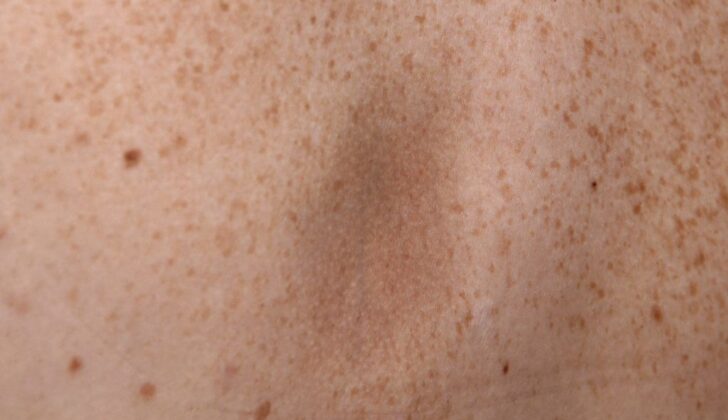
Atrophoderma of Pasini and Pierini is a condition that gradually develops over time. Initially, it might not present any symptoms. The first signs usually appear as skin patches on the back, but can also spread to the chest, arms, and stomach area. The patches can vary greatly in size, from tiny markings a few millimeters in diameter to larger ones spanning several centimeters. These patches sit slightly below the surrounding skin and have sharp edges, which gives them a unique ‘cliff-drop’ appearance. They are typically darker than the surrounding skin, but some people may have skin-colored or lighter patches.
Important to note is that these skin patches do not show any signs of inflammation, and when touched, they do not feel hard or swollen. The surrounding skin generally looks normal with no apparent changes. The patches can occur on both sides of the body. However, there have been instances where they only appeared on one side.
- Initial signs appear on the back
- Can spread to chest, arms, and stomach
- Patches vary from small markings to larger ones
- Patches sit slightly below the surrounding skin
- Patches have a unique ‘cliff-drop’ appearance
- Typically darker than the surrounding skin, but can also be skin-colored or lighter
- No signs of inflammation or hardness in the patches
- Surrounding skin looks normal
- Patches can occur on both or just one side of the body
Testing for Atrophoderma of Pasini and Pierini
In simple terms, a specific type of test called an enzyme-linked immunosorbent assay is used to detect antibodies created by the body in response to B. burgdorferi, a bacterium linked with certain diseases.
Doctors may also utilize specific imaging tools such as magnetic resonance imaging or a particular type of ultrasound known as 13-MHz B-mode ultrasonography to measure the thickness of the lesion and the skin around it. This is done to document any thinning of the skin in the area of the lesion. However, this is usually not necessary.
On the microscopic level, the changes to the skin are minimal but significant. They show that the skin’s volume is reduced compared to normal skin. Other characteristics that may be observed include a certain level of uniform texture and clumping of a protein called collagen in the deeper layers of the skin, some swelling in the spaces between cells, and a slight gathering of specific immune cells. The sweat glands, hair follicle-related structures, and skin-related structures usually appear normal. The layer of skin on top, known as the epidermis, is typically normal but may show slight thinning with flattened structures called rete ridges.
Treatment Options for Atrophoderma of Pasini and Pierini
Unfortunately, there isn’t a one-size-fits-all treatment for atrophoderma of Pasini and Pierini, a skin condition that causes changes in the skin’s color and texture.
Success has been seen in some patients who tested positive for the B. burgdorferi antibody, and were treated with tetracyclines like doxycycline. This antibiotic, taken at a dose of 200 mg per day, seems to help prevent the formation of new skin lesions. That said, the results with this treatment approach have been mixed, so it’s not considered a definitive solution.
In a case study of 25 patients, who were given either oral penicillin or tetracycline for a period of 2
to 3 weeks, 20 showed improvements in their skin condition. Interestingly, 4 out of the 6 patients who didn’t get any treatment did not show any disease progression.
In addition to these treatments, some topical medications like corticosteroids, antimalarial drugs including hydroxychloroquine, and calcineurin inhibitors, have also been used. These treatments seem to work especially well in patients whose condition is associated with lupus. However, like other treatments, responses can be inconsistent.
Lastly, one approach that has shown promise at improving the darker skin patches associated with this condition is treatment with a specific type of laser called a Q-switched Alexandrite laser.
What else can Atrophoderma of Pasini and Pierini be?
Here is a list of some medical conditions that dermatologists often come across:
- Atrophoderma of Moulin
- Anetoderma
- Lichen Sclerosis
- Morphea
- Postinflammatory Hyperpigmentation (a condition where the skin becomes darker after an inflammatory disease)
It’s important to consult a healthcare professional who can correctly diagnose these conditions based on your symptoms and medical history.
What to expect with Atrophoderma of Pasini and Pierini
The disease usually worsens over a period of 10 to 20 years. After this, it generally stabilizes and doesn’t get worse, but the existing skin abnormalities don’t heal or disappear. Up until 1982, no cases were reported of this disease developing into a more serious condition known as systemic sclerosis.
However, in 1982, a team led by Bisaccia reported a unique case. They detailed the experience of a 42-year-old woman who initially had typical symptoms of this disease on her back. Unfortunately, she developed a more severe condition called systemic sclerosis, which included symptoms like Raynaud’s phenomenon (where fingers and toes feel numb, prickly and cold in response to cold temperatures or stress), sclerodactyly (a hardening and tightening of the skin of the fingers or toes), and pulmonary fibrosis (a lung disease that occurs when lung tissue becomes damaged and scarred).


